Antibody data
- Antibody Data
- Antigen structure
- References [2]
- Comments [0]
- Validations
- Western blot [9]
- Immunocytochemistry [1]
- Immunohistochemistry [1]
Submit
Validation data
Reference
Comment
Report error
- Product number
- GTX100467 - Provider product page

- Provider
- GeneTex
- Proper citation
- GeneTex Cat#GTX100467, RRID:AB_11175804
- Product name
- uPAR antibody
- Antibody type
- Polyclonal
- Reactivity
- Human
- Host
- Rabbit
Submitted references Effects of TGF-β1 on plasminogen activation in human dental pulp cells: Role of ALK5/Smad2, TAK1 and MEK/ERK signalling.
Characterization and mechanism of stress-induced translocation of 78-kilodalton glucose-regulated protein (GRP78) to the cell surface.
Chang MC, Chang HH, Lin PS, Huang YA, Chan CP, Tsai YL, Lee SY, Jeng PY, Kuo HY, Yeung SY, Jeng JH
Journal of tissue engineering and regenerative medicine 2018 Apr;12(4):854-863
Journal of tissue engineering and regenerative medicine 2018 Apr;12(4):854-863
Characterization and mechanism of stress-induced translocation of 78-kilodalton glucose-regulated protein (GRP78) to the cell surface.
Tsai YL, Zhang Y, Tseng CC, Stanciauskas R, Pinaud F, Lee AS
The Journal of biological chemistry 2015 Mar 27;290(13):8049-64
The Journal of biological chemistry 2015 Mar 27;290(13):8049-64
No comments: Submit comment
Supportive validation
- Submitted by
- GeneTex (provider)
- Main image

- Experimental details
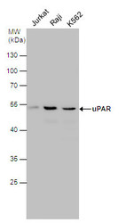
- uPAR antibody detects PLAUR protein by Western blot analysis.A. 30 µg Jurkat whole cell lysate/extractB. 30 µg Raji whole cell lysate/extractC. 30 µg K562 whole cell lysate/extract10 % SDS-PAGEuPAR antibody (GTX100467) dilution: 1:1000
- Validation comment
- WB
- Submitted by
- GeneTex (provider)
- Main image

- Experimental details
- uPAR antibody detects uPAR protein by western blot analysis. Various whole cell extracts (30 £gg) were separated by 10% SDS-PAGE, and the membrane was blotted with uPAR antibody (GTX100467) diluted at 1:1000.
- Submitted by
- GeneTex (provider)
- Main image

- Experimental details
- uPAR antibody detects uPAR protein by western blot analysis. Various whole cell extracts (30 £gg) were separated by 10% SDS-PAGE, and the membrane was blotted with uPAR antibody (GTX100467) diluted at 1:1000.
- Submitted by
- GeneTex (provider)
- Main image

- Experimental details
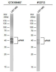
- Whole cell extract (30 ?g) was separated by 12% SDS-PAGE, and the membranes were blotted with uPAR antibody (GTX100467) diluted at 1:500 and competitor's antibody (CST#12713) diluted at 1:200. The HRP-conjugated anti-rabbit IgG antibody (GTX213110-01) was used to detect the primary antibody.
- Submitted by
- GeneTex (provider)
- Main image

- Experimental details
- Whole cell extract (30 ?g) was separated by 12% SDS-PAGE, and the membranes were blotted with uPAR antibody (GTX100467) diluted at 1:500 and competitor's antibody (CST#12713) diluted at 1:200. The HRP-conjugated anti-rabbit IgG antibody (GTX213110-01) was used to detect the primary antibody.
- Submitted by
- GeneTex (provider)
- Main image

- Experimental details
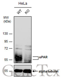
- Wild-type (WT) and uPAR knockout (KO) HeLa cell extracts (30 ?g) were separated by 10% SDS-PAGE, and the membrane was blotted with uPAR antibody (GTX100467) diluted at 1:500. The HRP-conjugated anti-rabbit IgG antibody (GTX213110-01) was used to detect the primary antibody, and the signal was developed with Trident ECL plus-Enhanced.
- Submitted by
- GeneTex (provider)
- Main image

- Experimental details
- Whole cell extract (30 ?g) was separated by 12% SDS-PAGE, and the membrane was blotted with uPAR antibody (GTX100467) diluted at 1:500.
- Submitted by
- GeneTex (provider)
- Main image

- Experimental details
- Whole cell extract (30 ?g) was separated by 12% SDS-PAGE, and the membrane was blotted with uPAR antibody (GTX100467) diluted at 1:1000. The HRP-conjugated anti-rabbit IgG antibody (GTX213110-01) was used to detect the primary antibody.
- Submitted by
- GeneTex (provider)
- Main image

- Experimental details
- Wild-type (WT) and uPAR knockout (KO) HeLa cell extracts (30 ?g) were separated by 10% SDS-PAGE, and the membrane was blotted with uPAR antibody (GTX100467) diluted at 1:500. The HRP-conjugated anti-rabbit IgG antibody (GTX213110-01) was used to detect the primary antibody, and the signal was developed with Trident ECL plus-Enhanced.
Supportive validation
- Submitted by
- GeneTex (provider)
- Main image

- Experimental details
- uPAR antibody detects uPAR protein at cytoplasm by immunofluorescent analysis.Sample: A431 cells were fixed in 4% paraformaldehyde at RT for 15 min.Green: uPAR protein stained by uPAR antibody (GTX100467) diluted at 1:500.Blue: Hoechst 33342 staining.
Supportive validation
- Submitted by
- GeneTex (provider)
- Main image

- Experimental details
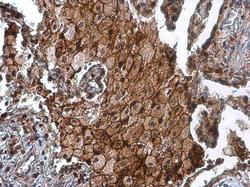
- uPAR antibody detects uPAR protein at cell membrane by immunohistochemical analysis.Sample: Paraffin-embedded human lung cancer.uPAR stained by uPAR antibody (GTX100467) diluted at 1:500.
 Explore
 Validate
 Learn
Learn Western blot