 Explore
 Validate
 Learn
Learn Western blot
Antibody data
- Antibody Data
- Antigen structure
- References [3]
- Comments [0]
- Validations
- Western blot [9]
- Immunocytochemistry [4]
- Immunohistochemistry [4]
- Flow cytometry [2]
Submit
Validation data
Reference
Comment
Report error
- Product number
- MA5-17029 - Provider product page

- Provider
- Invitrogen Antibodies
- Product name
- ALDH2 Monoclonal Antibody (4G6A3)
- Antibody type
- Monoclonal
- Antigen
- Purifed from natural sources
- Description
- MA5-17029 targets ALDH2 in FACS, ICC, IHC, IF and WB applications and shows reactivity with Human, Mouse, and Rat samples.
- Antibody clone number
- 4G6A3
- Concentration
- 1 mg/mL
Submitted references A role for aldehyde dehydrogenase (ALDH) 2 in angiotensin II-mediated decrease in angiogenesis of coronary endothelial cells.
Solid-phase inclusion as a mechanism for regulating unfolded proteins in the mitochondrial matrix.
AAV Gene Therapy for Alcoholism: Inhibition of Mitochondrial Aldehyde Dehydrogenase Enzyme Expression in Hepatoma Cells.
Roy B, Palaniyandi SS
Microvascular research 2021 May;135:104133
Microvascular research 2021 May;135:104133
Solid-phase inclusion as a mechanism for regulating unfolded proteins in the mitochondrial matrix.
Ruan L, McNamara JT, Zhang X, Chang AC, Zhu J, Dong Y, Sun G, Peterson A, Na CH, Li R
Science advances 2020 Aug;6(32):eabc7288
Science advances 2020 Aug;6(32):eabc7288
AAV Gene Therapy for Alcoholism: Inhibition of Mitochondrial Aldehyde Dehydrogenase Enzyme Expression in Hepatoma Cells.
Sanchez AC, Li C, Andrews B, Asenjo JA, Samulski RJ
Human gene therapy 2017 Sep;28(9):717-725
Human gene therapy 2017 Sep;28(9):717-725
No comments: Submit comment
Supportive validation
- Submitted by
- Invitrogen Antibodies (provider)
- Main image

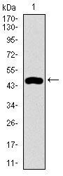
- Experimental details
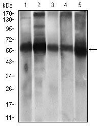
- Western blot analysis of ALDH2 in HEK293 (lane 1) and ALDH2-hIgGFc transfected HEK293 (lane 2) cell lysate using ALDH2 monoclonal antibody (Product # MA5-17029).

- Submitted by
- Invitrogen Antibodies (provider)
- Main image
- Experimental details
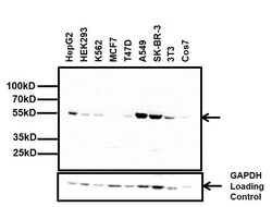
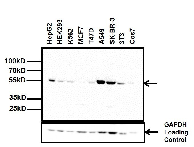
- Western blot analysis of ALDH2 was performed by loading 20 µg of the indicated whole cell lysates and 5 µL of PageRuler Plus Prestained Protein Ladder (Product # 26619) onto a 4-20% Tris-Glycine polyacrylamide gel (Product # WT4202BX10). Proteins were transferred to a nitrocellulose membrane using the G2 Blotter (Product # 62288), and blocked with 5% Milk in TBST for 1 hour at room temperature. ALDH2 was detected at ~56 kDa using a ALDH2 mouse monoclonal antibody (Product # MA5-17029) at a concentration of 1 µg/mL in blocking buffer overnight at 4°C on a rocking platform, followed by a Goat anti-mouse IgG (H+L) Superclonal™ Secondary Antibody, HRP conjugate (Product # A28177) at a dilution of 1:2000 for at least one hour at room temperature. Chemiluminescent detection was performed using SuperSignal West Pico (Product # 34078).
- Submitted by
- Invitrogen Antibodies (provider)
- Main image
- Experimental details
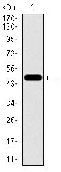
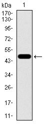
- Western blot analysis of ALDH2 using a ALDH2 monoclonal antibody (Product # MA5-17029) against a human ALDH2 recombinant protein.
- Submitted by
- Invitrogen Antibodies (provider)
- Main image

- Experimental details
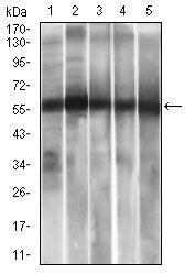
- Western blot analysis of ALDH2 using ALDH2 monoclonal antibody (Product # MA5-17029) in HepG2 (1), A549 (2) cell lysate, and rat liver (3), mouse liver (4), mouse brain (5) tissue lysate.
- Submitted by
- Invitrogen Antibodies (provider)
- Main image
- Experimental details
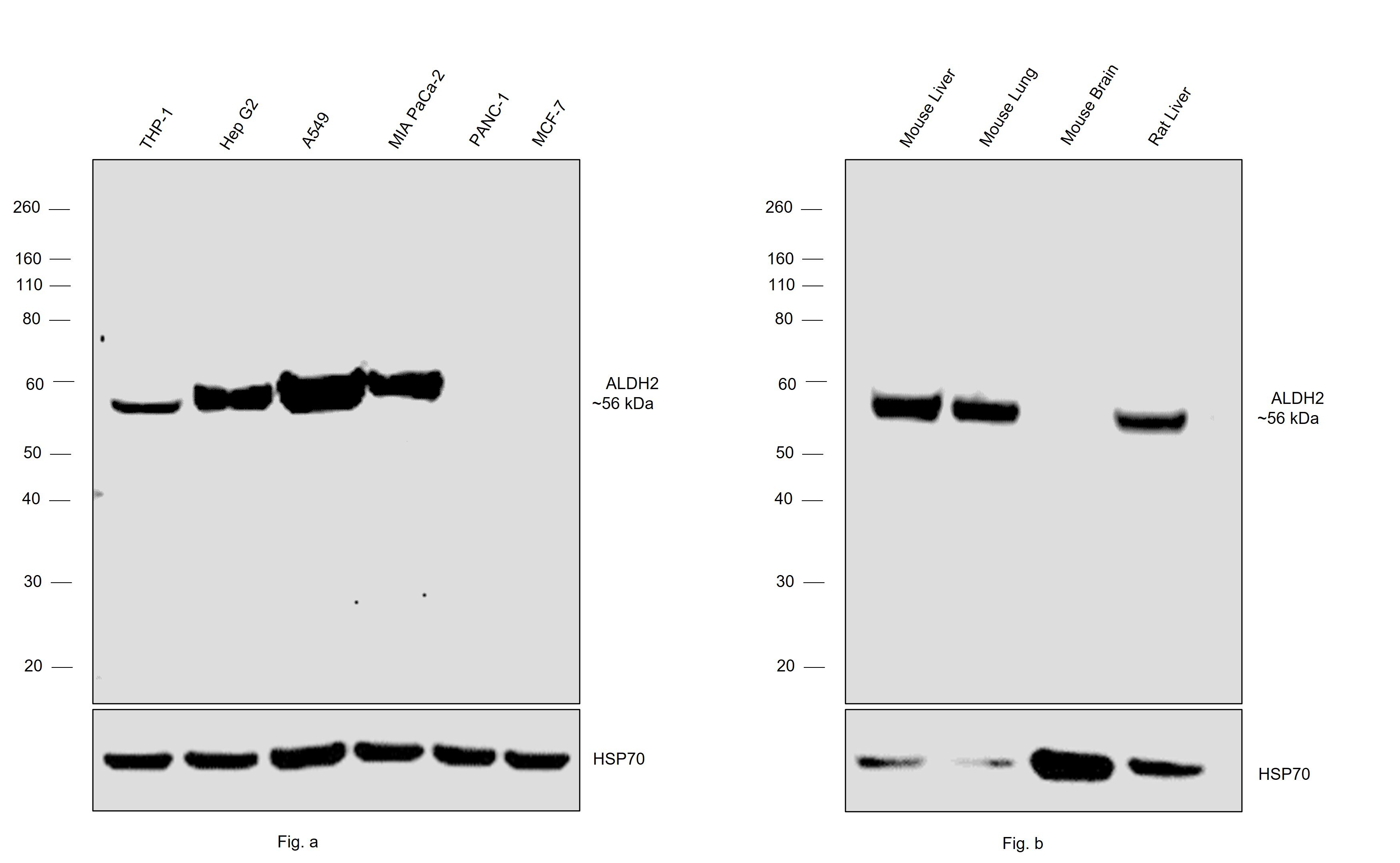
- Western blot was performed using Anti-ALDH2 Monoclonal Antibody (4G6A3) (Product # MA5-17029) and a 56 kDa band corresponding to acetaldehyde dehydrogenase 2; ALDH2 was observed across the panel tested except for PANC-1 and MCF7 (Fig.a) and Mouse Brain (Fig.b) which are reported to be low expressors of ALDH2. Whole cell extracts (30 µg lysate) of THP-1 (Lane 1), Hep G2 (Lane 2), A549 (Lane 3), MIA PaCa-2 (Lane 4), PANC-1 (Lane 5), MCF7 (Lane 6) (Fig.a) and Mouse Liver (Lane 1), Mouse Lung (Lane 2), Mouse Brain (Lane 3), Rat Liver (Lane 4) (Fig.b) were electrophoresed using NuPAGE™ 4-12% Bis-Tris Protein Gel (Product # NP0321BOX), 10 well. Resolved proteins were then transferred onto a nitrocellulose membrane (Product # IB23001) by iBlot® 2 Dry Blotting System (Product # IB21001). The blot was probed with the primary antibody (1 µg/mL dilution) and detected by chemiluminescence with Goat anti-Mouse IgG (H+L) Superclonal™ Recombinant Secondary Antibody, HRP (Product # A28177, 1:20,000 dilution) using the iBright™ FL1500 Imaging System (Product # A44115). Chemiluminescent detection was performed using SuperSignal™ West Pico PLUS Chemiluminescent Substrate (Product # 34580).
- Submitted by
- Invitrogen Antibodies (provider)
- Main image

- Experimental details
- Western blot analysis of ALDH2 in HepG2 (lane 1), A549 (lane 2) cell lysate, and rat liver (lane 3), mouse liver (lane 4), mouse brain (lane 5) tissue lysate using ALDH2 monoclonal antibody (Product # MA5-17029).
- Submitted by
- Invitrogen Antibodies (provider)
- Main image
- Experimental details
- Western blot analysis of ALDH2 monoclonal antibody (Product # MA5-17029) against human ALDH2 recombinant protein. Expected MW is 47.4 kDa.

- Submitted by
- Invitrogen Antibodies (provider)
- Main image

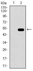
- Experimental details
- Western blot analysis of ALDH2 in HEK293 (lane 1) and ALDH2-hIgGFc transfected HEK293 (lane 2) cell lysate using ALDH2 monoclonal antibody (Product # MA5-17029).
- Submitted by
- Invitrogen Antibodies (provider)
- Main image
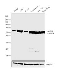
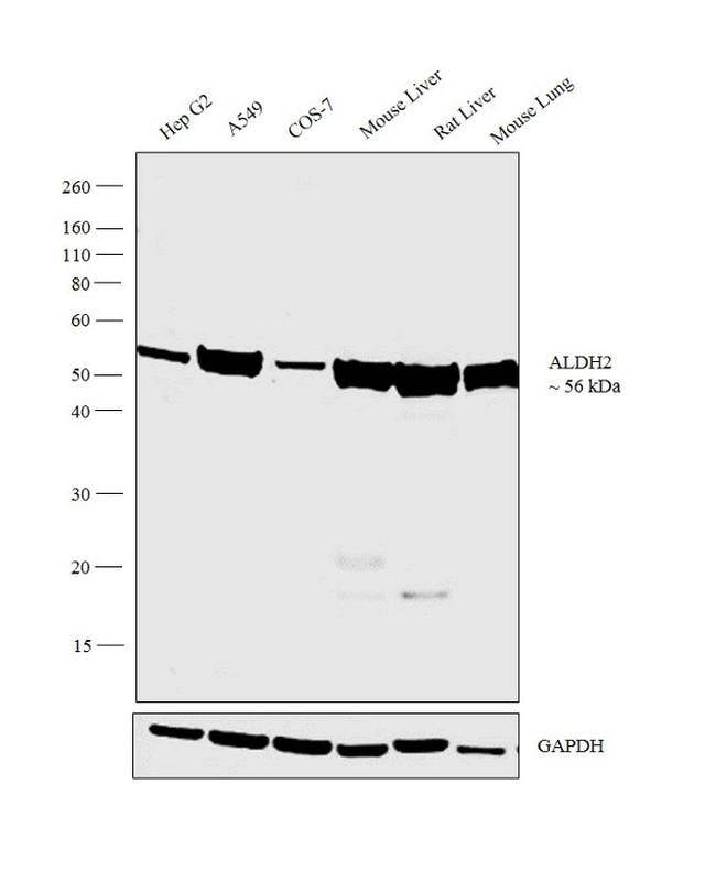
- Experimental details
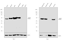
- Western blot analysis was performed on whole cell extracts (30 µg lysate) of Hep G2 (Lane 1), A549 (Lane 2), COS-7 (Lane 3), tissue extracts of Mouse Liver (Lane 4), Rat Liver (Lane 5) and Mouse Lung (Lane 6). The blot was probed with Anti-ALDH2 Monoclonal Antibody (Product # MA5-17029, 1µg/ml) and detected by chemiluminescence using Goat anti-Mouse IgG (H+L) Superclonal™ Secondary Antibody, HRP conjugate (Product # A28177, 0.25 µg/ml, 1:4000 dilution). A 56 kDa band corresponding to ALDH2 was observed across cell lines and tissues tested.
Supportive validation
- Submitted by
- Invitrogen Antibodies (provider)
- Main image

- Experimental details
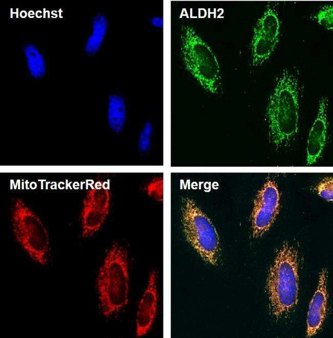
- Immunofluorescent analysis of ALDH2 (green) in HeLa cells. The cells were fixed with 4% Paraformaldehyde for 15 minutes, permeabilized with 0.1% Triton X-100 for 15 minutes, and blocked with 3% BSA for 30 minutes at room temperature. Cells were stained with an ALDH2 mouse monoclonal antibody (Product # MA5-17029) at a concentration of 1.25 µg/mL in blocking buffer for 1 hour at room temperature, and then incubated with a Goat anti-Mouse IgG (H+L) Secondary Antibody, Alexa Fluor Plus 488 conjugate (Product # A32723) at a dilution of 1:500 for at least 30 minutes at a room temperature in the dark (green). Nuclei (blue) were stained with Hoechst 33342 (Product # 62249). Mitochondria (red) was stained with MitoTracker® Red CMXRos (Product # M7512). Images were taken on a Thermo Scientific ToxInsight Instrument at 20X magnification.
- Submitted by
- Invitrogen Antibodies (provider)
- Main image

- Experimental details
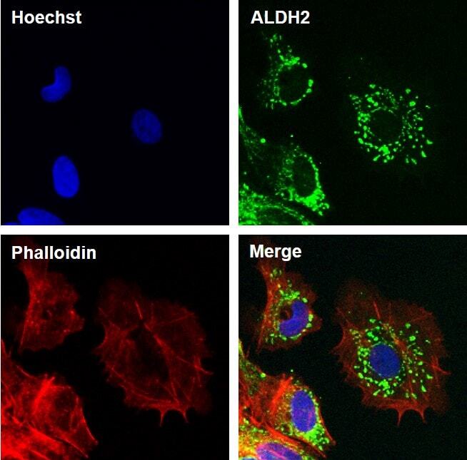
- Immunofluorescent analysis of ALDH2 (green) in HeLa cells. The cells were fixed with 4% Paraformaldehyde for 15 minutes, permeabilized with 0.1% Triton X-100 for 15 minutes, and blocked with 3% BSA for 30 minutes at room temperature. Cells were stained with an ALDH2 mouse monoclonal antibody (Product # MA5-17029) at a concentration of 2.5 µg/mL in blocking buffer for 1 hour at room temperature, and then incubated with a Goat anti-Mouse IgG (H+L) Secondary Antibody, Alexa Fluor Plus 488 conjugate (Product # A32723) at a dilution of 1:500 for at least 30 minutes at a room temperature in the dark (green). F-actin (red) was stained by Dylight 554 Phalloidin (Product #21834) and nuclei (blue) were stained with Hoechst 33342 (Product # 62249). Images were taken on a Thermo Scientific ToxInsight Instrument at 20X magnification.
- Submitted by
- Invitrogen Antibodies (provider)
- Main image
- Experimental details
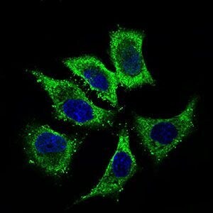
- Immunofluorescence analysis of HepG2 cells using ALDH2 monoclonal antibody (Product # MA5-17029) (Green). Blue: DRAQ5 fluorescent DNA dye. Red: actin filaments have been labeled with phalloidin.

- Submitted by
- Invitrogen Antibodies (provider)
- Main image

- Experimental details
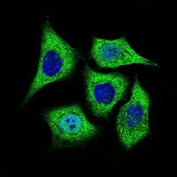
- Immunofluorescent analysis of HepG2 cells using ALDH2 monoclonal antibody (Product # MA5-17029) (green). Blue: fluorescent DNA dye.
Supportive validation
- Submitted by
- Invitrogen Antibodies (provider)
- Main image

- Experimental details
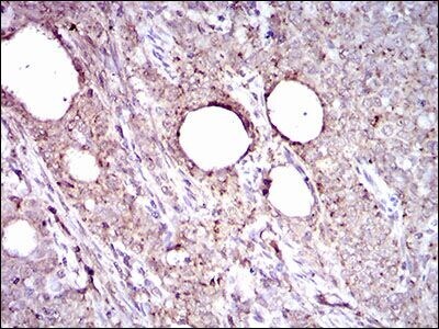
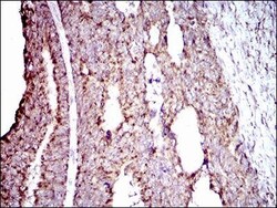
- Immunohistochemical analysis of paraffin-embedded cervical cancer tissues using ALDH2 monoclonal antibody (Product # MA5-17029) followed with DAB staining.
- Submitted by
- Invitrogen Antibodies (provider)
- Main image

- Experimental details
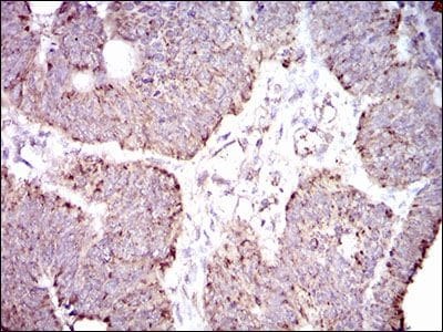
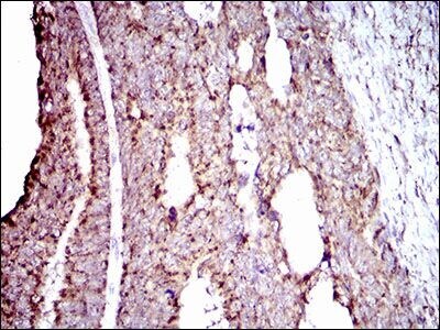
- Immunohistochemical analysis of paraffin-embedded rectum cancer tissues using ALDH2 monoclonal antibody (Product # MA5-17029) followed with DAB staining.
- Submitted by
- Invitrogen Antibodies (provider)
- Main image
- Experimental details
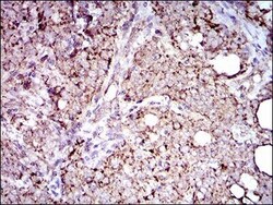
- Immunohistochemical analysis of paraffin-embedded cervical cancer tissues using ALDH2 monoclonal antibody (Product # MA5-17029) with DAB staining.
- Submitted by
- Invitrogen Antibodies (provider)
- Main image
- Experimental details
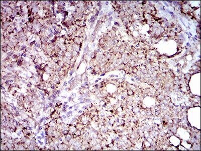
- Immunohistochemical analysis of paraffin-embedded rectum cancer tissues using ALDH2 monoclonal antibody (Product # MA5-17029) with DAB staining.
Supportive validation
- Submitted by
- Invitrogen Antibodies (provider)
- Main image
- Experimental details
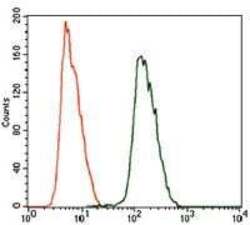
- Flow cytometric analysis of HeLa cells using ALDH2 monoclonal antibody (Product # MA5-17029) (green) and negative control (purple).
- Submitted by
- Invitrogen Antibodies (provider)
- Main image
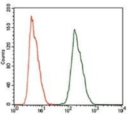
- Experimental details
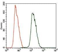
- Flow cytometric analysis of HeLa cells using ALDH2 monoclonal antibody (Product # MA5-17029) (green) and negative control (red).
