 Explore
 Validate
 Learn
Learn Western blot
Antibody data
- Antibody Data
- Antigen structure
- References [0]
- Comments [0]
- Validations
- Western blot [2]
- Immunocytochemistry [1]
- Flow cytometry [1]
Submit
Validation data
Reference
Comment
Report error
- Product number
- MA5-37560 - Provider product page

- Provider
- Invitrogen Antibodies
- Product name
- RAB23 Monoclonal Antibody (427CT2.1.1)
- Antibody type
- Monoclonal
- Antigen
- Purifed from natural sources
- Reactivity
- Human, Mouse
- Host
- Mouse
- Isotype
- IgG
- Antibody clone number
- 427CT2.1.1
- Vial size
- 400 µL
- Concentration
- 0.5 mg/mL
- Storage
- Store at 4°C short term. For long term storage, store at -20°C, avoiding freeze/thaw cycles.
No comments: Submit comment
Supportive validation
- Submitted by
- Invitrogen Antibodies (provider)
- Main image
- Experimental details
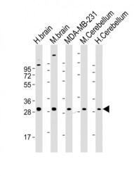
- Western Blot analysis of RAB23 using the RAB23 Monoclonal Antibody (Product # MA5-37560) at a 1:2000 dilution. Lane 1: human brain lysate, Lane 2: mouse brain lysate, Lane 3: MDA-MB-231 whole cell lysate, Lane 4: mouse Cerebellum lysate, Lane 5: human Cerebellum lysate. Lysates/proteins at 20 µg per lane. Secondary Goat Anti-mouse IgG, (H+L), Peroxidase conjugated at 1:10,000 dilution. Predicted band size : 27 kDa. Blocking/Dilution buffer: 5% NFDM/TBST.

- Submitted by
- Invitrogen Antibodies (provider)
- Main image

- Experimental details
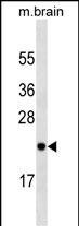
- Western Blot analysis of RAB23 using the RAB23 Monoclonal Antibody (Product # MA5-37560) in mouse brain tissue lysates (35 µg/lane).
Supportive validation
- Submitted by
- Invitrogen Antibodies (provider)
- Main image

- Experimental details
- Immunocytochemistry/Immunofluorescent analysis of RAB23 in 4% paraformaldehyde-fixed, 0.1% Triton X-100 permeabilized U-2 OS cells using RAB23 Monoclonal Antibody (Product # MA5-37560) at a 1:25 dilution, followed by Dylight® 488-conjugated goat anti-mouse IgG (174309) secondary antibody at 1:200 dilution (green). Immunofluorescence image showing cytoplasm staining on U-2 OS cell line. Cytoplasmic actin is detected with Dylight® 554 Phalloidin at 1:100 dilution (red).The nuclear counter stain is DAPI (blue).

Supportive validation
- Submitted by
- Invitrogen Antibodies (provider)
- Main image

- Experimental details
- Flow Cytometry analysis showing U-2 OS cells stained with RAB23 Monoclonal Antibody (Product # MA5-37560) at a 1:25 dilution (green line). The cells were fixed with 2% paraformaldehyde (10 min) and then permeabilized with 90% methanol for 10 min. The cells were then incubated in 2% BSA to block non-specific protein-protein interactions followed by the RAB23 Monoclonal Antibody for 60 min at 37°C. The secondary antibody used was Goat-Anti-Mouse IgG, DyLight® 488 Conjugated Highly Cross-Adsorbed at 1:200 dilution for 40 min at 37°C. Isotype control antibody (blue line) was mouse IgG1(1µg/1x10^6 cells) used under the same conditions. Acquisition of >10, 000 events was performed.
