 Explore
 Validate
 Learn
Learn Western blot
 Immunohistochemistry
Antibody data
- Antibody Data
- Antigen structure
- References [2]
- Comments [0]
- Validations
- Immunohistochemistry [1]
Submit
Validation data
Reference
Comment
Report error
- Product number
- AF2784 - Provider product page

- Provider
- Novus Biologicals
- Product name
- Goat Polyclonal CLEC4F/CLECSF13 Antibody
- Antibody type
- Polyclonal
- Description
- Antigen Affinity-purified. Detects mouse CLEC4F/CLECSF13 in direct ELISAs and Western blots.
- Reactivity
- Mouse
- Host
- Goat
- Conjugate
- Unconjugated
- Isotype
- IgG
- Vial size
- 100 ug
- Concentration
- LYOPH
- Storage
- Use a manual defrost freezer and avoid repeated freeze-thaw cycles. 12 months from date of receipt, -20 to -70 degreesC as supplied. 1 month, 2 to 8 degreesC under sterile conditions after reconstitution. 6 months, -20 to -70 degreesC under sterile conditions after reconstitution.
Submitted references Characterizing responsive and refractory orthotopic mouse models of hepatocellular carcinoma in cancer immunotherapy.
Growth differentiation factor 15 ameliorates nonalcoholic steatohepatitis and related metabolic disorders in mice.
Hage C, Hoves S, Ashoff M, Schandl V, Hört S, Rieder N, Heichinger C, Berrera M, Ries CH, Kiessling F, Pöschinger T
PloS one 2019;14(7):e0219517
PloS one 2019;14(7):e0219517
Growth differentiation factor 15 ameliorates nonalcoholic steatohepatitis and related metabolic disorders in mice.
Kim KH, Kim SH, Han DH, Jo YS, Lee YH, Lee MS
Scientific reports 2018 May 1;8(1):6789
Scientific reports 2018 May 1;8(1):6789
No comments: Submit comment
Supportive validation
- Submitted by
- Novus Biologicals (provider)
- Main image
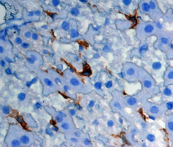
- Experimental details
- CLEC4F/CLECSF13 in Mouse Liver. CLEC4F/CLECSF13 was detected in perfusion fixed frozen sections of mouse liver using Goat Anti-Mouse CLEC4F/CLECSF13 Antigen Affinity-purified Polyclonal Antibody (Catalog # AF2784) at 5 µg/mL overnight at 4 °C. Tissue was stained using the Anti-Goat HRP-DAB Cell & Tissue Staining Kit (brown; Catalog # CTS008) and counterstained with hematoxylin (blue). Specific staining was localized to Kupffer cells. View our protocol for Chromogenic IHC Staining of Frozen Tissue Sections.
