Explore
 Validate
 Learn
Learn Western blot
Antibody data
- Antibody Data
- Antigen structure
- References [1]
- Comments [0]
- Validations
- Western blot [3]
- Immunohistochemistry [1]
- Other assay [1]
Submit
Validation data
Reference
Comment
Report error
- Product number
- PA5-31090 - Provider product page

- Provider
- Invitrogen Antibodies
- Product name
- OAS3 Polyclonal Antibody
- Antibody type
- Polyclonal
- Antigen
- Recombinant protein fragment
- Description
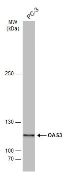
- Recommended positive controls: PC-3.
- Concentration
- 0.33 mg/mL
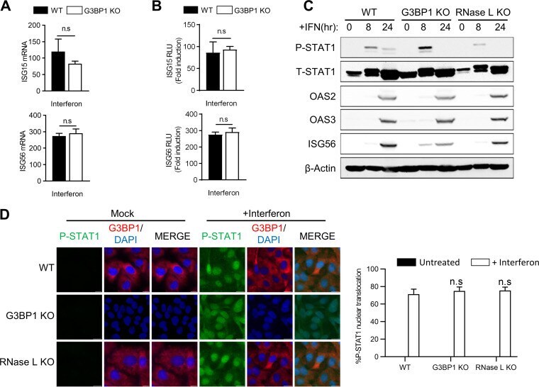
Submitted references RNase L Amplifies Interferon Signaling by Inducing Protein Kinase R-Mediated Antiviral Stress Granules.
Manivannan P, Siddiqui MA, Malathi K
Journal of virology 2020 Jun 16;94(13)
Journal of virology 2020 Jun 16;94(13)
No comments: Submit comment
Supportive validation
- Submitted by
- Invitrogen Antibodies (provider)
- Main image
- Experimental details
- Western blot analysis of OAS3 using 30 µg of A431 lysate. Samples were loaded onto a 7.5% SDS-PAGE gel and probed with an OAS3 polyclonal antibody (Product # PA5-31090) at a dilution of 1:3000.

- Submitted by
- Invitrogen Antibodies (provider)
- Main image

- Experimental details
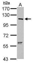
- Western blot analysis of OAS3 was performed by separating 30 µg of whole cell extract by 5% SDS-PAGE. Proteins were transferred to a membrane and probed with a OAS3 Polyclonal Antibody (Product # PA5-31090) at a dilution of 1:3000. The HRP-conjugated anti-rabbit IgG antibody was used to detect the primary antibody.
- Submitted by
- Invitrogen Antibodies (provider)
- Main image
- Experimental details
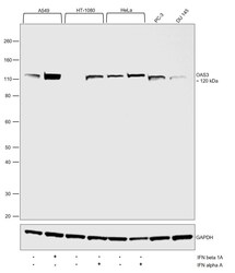
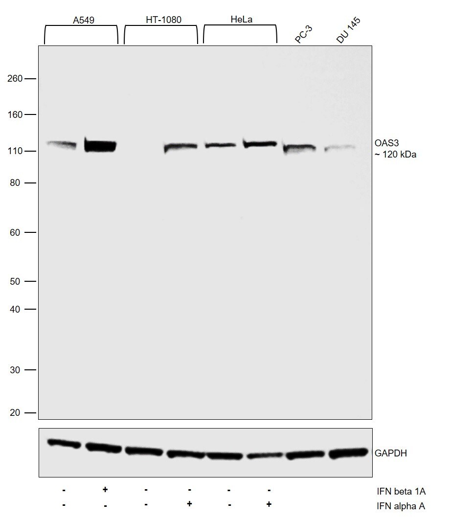
- Western Blot was performed using Anti-OAS3 Polyclonal Antibody (Product # PA5-31090) and a 120 kDa band corresponding to 2-5-oligoadenylate synthase 3 was observed. Whole cell extracts (30 µg lysate) of A549 (Lane 1), A549 treated with IFN beta 1A (1000 U/mL, O/N) (Lane 2), HT-1080 (Lane 3), HT-1080 treated with IFN alpha A (2000 U/mL, O/N) (Lane 4), HeLa (Lane 5), HeLa treated with IFN alpha A (10 ng/mL,16 hrs) (Lane 6), PC-3 (Lane 7) and DU 145 (Lane 8) were electrophoresed using NuPAGE™ 4-12% Bis-Tris Protein Gel (Product # NP0322BOX). Resolved proteins were then transferred onto a nitrocellulose membrane (Product # IB23001) by iBlot® 2 Dry Blotting System (Product # IB21001). The blot was probed with the primary antibody (1:2000) and detected by chemiluminescence with Goat anti-Rabbit IgG (H+L) Superclonal™ Recombinant Secondary Antibody, HRP (Product # A27036, 1:4000) using the iBright FL 1000 (Product # A32752). Chemiluminescent detection was performed using Novex® ECL Chemiluminescent Substrate Reagent Kit (Product # WP20005). Upregulation in the expression of OAS3 was observed upon Interferon treatment in all the models tested (https://doi.org/10.1073/pnas.1519657113).
Supportive validation
- Submitted by
- Invitrogen Antibodies (provider)
- Main image
- Experimental details
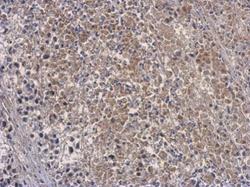
- Immunohistochemical analysis of paraffin-embedded D54MG xenograft, using OAS3 (Product # PA5-31090) antibody at 1:500 dilution. Antigen Retrieval: EDTA based buffer, pH 8.0, 15 min.
Supportive validation
- Submitted by
- Invitrogen Antibodies (provider)
- Main image
- Experimental details
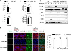
- FIG 5 Effect of avSG formation on IFN signaling. (A and B) HT1080 WT and G3BP1 KO cells were treated with IFN-beta (1,000 U/mL) for 24 h, and ISG15 and ISG56 mRNA levels were measured and normalized to those of GAPDH by qRT-PCR (A), or the cells were transfected with ISG15-luc or ISG56-luc reporter constructs, along with beta-galactosidase plasmids, and 24 h later treated with IFN-beta (1,000 U/mL), and luciferase activity was measured and normalized to beta-galactosidase levels (B). (C) WT, G3BP1 KO, and RNase L KO cells were treated with IFN-beta (1,000 U/mL) for the indicated times, and cell lysates were analyzed for phosphorylation of STAT1 and induction of OAS2, OAS3, and ISG56 in immunoblots. beta-Actin was used to normalize loading. (D) WT, G3BP1 KO, and RNase L KO cells were treated with IFN-beta (1,000 U/mL) for 16 h, and nuclear translocation of P-STAT1 was determined by immunofluorescence; nuclei were stained with DAPI. (Right) Quantification of P-STAT1 nuclear translocation from five random fields. The data represent means + SE for the results of three independent experiments. n.s, not significant.