Explore
 Validate
 Learn
Learn Western blot
 Immunoprecipitation
Antibody data
- Antibody Data
- Antigen structure
- References [3]
- Comments [0]
- Validations
- Western blot [2]
- Immunohistochemistry [1]
Submit
Validation data
Reference
Comment
Report error
- Product number
- AF1513 - Provider product page

- Provider
- Novus Biologicals
- Product name
- Goat Polyclonal ACE/CD143 Antibody
- Antibody type
- Polyclonal
- Description
- Antigen Affinity-purified. Detects mouse ACE in direct ELISAs and Western blots. In direct ELISAs, approximately 20% cross-reactivity with recombinant human ACE is observed.
- Reactivity
- Mouse
- Host
- Goat
- Conjugate
- Unconjugated
- Isotype
- IgG
- Vial size
- 100 ug
- Concentration
- LYOPH
- Storage
- Use a manual defrost freezer and avoid repeated freeze-thaw cycles. 12 months from date of receipt, -20 to -70 degreesC as supplied. 1 month, 2 to 8 degreesC under sterile conditions after reconstitution. 6 months, -20 to -70 degreesC under sterile conditions after reconstitution.
Submitted references ACE2 links amino acid malnutrition to microbial ecology and intestinal inflammation.
Major role for ACE-independent intrarenal ANG II formation in type II diabetes.
Angiotensin-converting enzyme 2 protects from severe acute lung failure.
Hashimoto T, Perlot T, Rehman A, Trichereau J, Ishiguro H, Paolino M, Sigl V, Hanada T, Hanada R, Lipinski S, Wild B, Camargo SM, Singer D, Richter A, Kuba K, Fukamizu A, Schreiber S, Clevers H, Verrey F, Rosenstiel P, Penninger JM
Nature 2012 Jul 25;487(7408):477-81
Nature 2012 Jul 25;487(7408):477-81
Major role for ACE-independent intrarenal ANG II formation in type II diabetes.
Park S, Bivona BJ, Kobori H, Seth DM, Chappell MC, Lazartigues E, Harrison-Bernard LM
American journal of physiology. Renal physiology 2010 Jan;298(1):F37-48
American journal of physiology. Renal physiology 2010 Jan;298(1):F37-48
Angiotensin-converting enzyme 2 protects from severe acute lung failure.
Imai Y, Kuba K, Rao S, Huan Y, Guo F, Guan B, Yang P, Sarao R, Wada T, Leong-Poi H, Crackower MA, Fukamizu A, Hui CC, Hein L, Uhlig S, Slutsky AS, Jiang C, Penninger JM
Nature 2005 Jul 7;436(7047):112-6
Nature 2005 Jul 7;436(7047):112-6
No comments: Submit comment
Supportive validation
- Submitted by
- Novus Biologicals (provider)
- Main image
- Experimental details
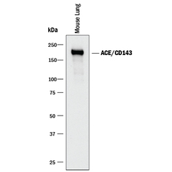
- Detection of Mouse ACE/CD143 by Western Blot. Western blot shows lysates of mouse lung tissue. PVDF membrane was probed with 0.05 µg/mL of Goat Anti-Mouse ACE/CD143 Antigen Affinity-purified Polyclonal Antibody (Catalog # AF1513) followed by HRP-conjugated Anti-Goat IgG Secondary Antibody (Catalog # HAF017). A specific band was detected for ACE/CD143 at approximately 180 kDa (as indicated). This experiment was conducted under reducing conditions and using Immunoblot Buffer Group 1.

- Submitted by
- Novus Biologicals (provider)
- Main image
- Experimental details
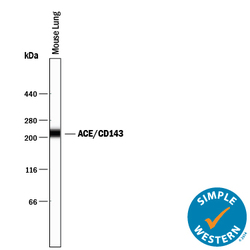
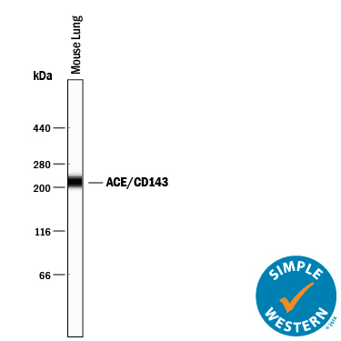
- Detection of Mouse ACE/CD143 by Simple WesternTM. Simple Western lane view shows lysates of mouse lung tissue, loaded at 0.2 mg/mL. A specific band was detected for ACE/CD143 at approximately 218 kDa (as indicated) using 10 µg/mL of Goat Anti-Mouse ACE/CD143 Antigen Affinity-purified Polyclonal Antibody (Catalog # AF1513) followed by 1:50 dilution of HRP-conjugated Anti-Goat IgG Secondary Antibody (Catalog # HAF109). This experiment was conducted under reducing conditions and using the 66-440 kDa separation system.
Supportive validation
- Submitted by
- Novus Biologicals (provider)
- Main image
- Experimental details
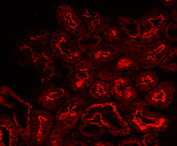
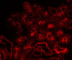
- ACE/CD143 in Mouse Kidney. ACE/CD143 was detected in perfusion fixed frozen sections of mouse kidney using 15 µg/mL Goat Anti-Mouse ACE/CD143 Antigen Affinity-purified Polyclonal Antibody (Catalog # AF1513) overnight at 4 °C. Tissue was stained (red). View our protocol for Fluorescent IHC Staining of Frozen Tissue Sections.