Explore
 Validate
 Learn
Learn Western blot
 ELISA
ELISAAntibody data
- Antibody Data
- Antigen structure
- References [0]
- Comments [0]
- Validations
- Western blot [1]
- Immunocytochemistry [4]
- Immunohistochemistry [8]
- Flow cytometry [1]
Submit
Validation data
Reference
Comment
Report error
- Product number
- RQ8169 - Provider product page

- Provider
- NSJ Bioreagents
- Product name
- VDAC1 Antibody
- Antibody type
- Polyclonal
- Description
- Antigen affinity purified
- Reactivity
- Human, Mouse, Rat
- Host
- Rabbit
- Vial size
- 100 µg
- Concentration
- 0.5mg/ml if reconstituted with 0.2ml sterile DI water
- Storage
- After reconstitution, the VDAC1 antibody can be stored for up to one month at 4oC. For long-term, aliquot and store at -20oC. Avoid repeated freezing and thawing.
No comments: Submit comment
Supportive validation
- Submitted by
- NSJ Bioreagents (provider)
- Main image

- Experimental details
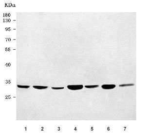
- Western blot testing of 1) human HeLa, 2) human A431, 3) human 293T, 4) rat heart, 5) rat liver, 6) mouse heart and 7) mouse liver tissue lysate with VDAC1 antibody. Predicted molecular weight 30~35 kDa.
Supportive validation
- Submitted by
- NSJ Bioreagents (provider)
- Main image

- Experimental details
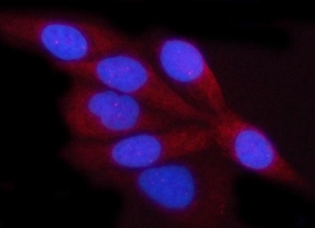
- Immunofluorescent staining of FFPE human HeLa cells with VDAC1 antibody (red) and DAPI nuclear stain (blue). HIER: steam section in pH6 citrate buffer for 20 min.
- Submitted by
- NSJ Bioreagents (provider)
- Main image

- Experimental details
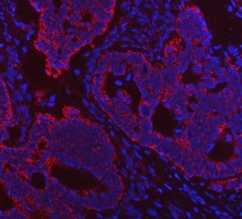
- Immunofluorescent staining of FFPE human intestinal cancer tissue with VDAC1 antibody (red) and DAPI nuclear stain (blue). HIER: steam section in pH8 EDTA buffer for 20 min.
- Submitted by
- NSJ Bioreagents (provider)
- Main image
- Experimental details
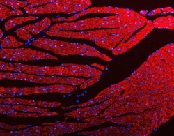
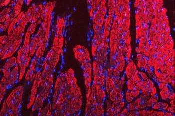
- Immunofluorescent staining of FFPE mouse heart tissue with VDAC1 antibody (red) and DAPI nuclear stain (blue). HIER: steam section in pH8 EDTA buffer for 20 min.

- Submitted by
- NSJ Bioreagents (provider)
- Main image

- Experimental details
- Immunofluorescent staining of FFPE rat heart tissue with VDAC1 antibody (red) and DAPI nuclear stain (blue). HIER: steam section in pH8 EDTA buffer for 20 min.
Supportive validation
- Submitted by
- NSJ Bioreagents (provider)
- Main image
- Experimental details
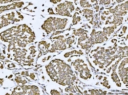
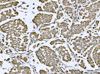
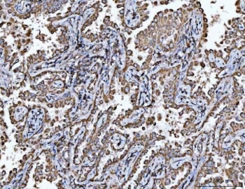
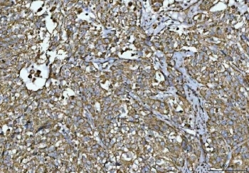
- IHC staining of FFPE human breast cancer tissue with VDAC1 antibody. HIER: boil tissue sections in pH8 EDTA for 20 min and allow to cool before testing.
- Submitted by
- NSJ Bioreagents (provider)
- Main image

- Experimental details
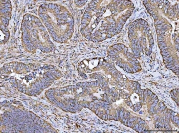
- IHC staining of FFPE human colorectal adenocarcinoma tissue with VDAC1 antibody. HIER: boil tissue sections in pH8 EDTA for 20 min and allow to cool before testing.
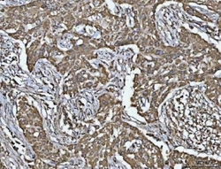
- Submitted by
- NSJ Bioreagents (provider)
- Main image
- Experimental details
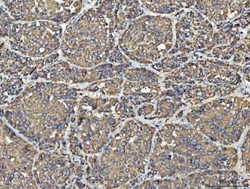
- IHC staining of FFPE human liver cancer tissue with VDAC1 antibody. HIER: boil tissue sections in pH8 EDTA for 20 min and allow to cool before testing.
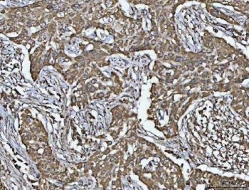
- Submitted by
- NSJ Bioreagents (provider)
- Main image
- Experimental details
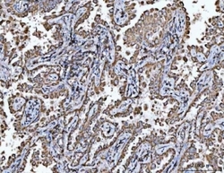
- IHC staining of FFPE human lung cancer tissue with VDAC1 antibody. HIER: boil tissue sections in pH8 EDTA for 20 min and allow to cool before testing.
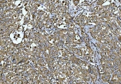
- Submitted by
- NSJ Bioreagents (provider)
- Main image
- Experimental details
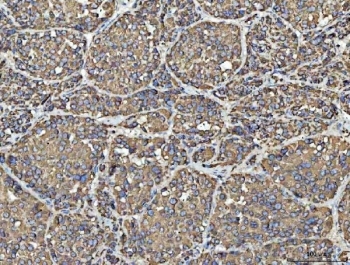
- IHC staining of FFPE human lymphadenoma tissue with VDAC1 antibody. HIER: boil tissue sections in pH8 EDTA for 20 min and allow to cool before testing.
- Submitted by
- NSJ Bioreagents (provider)
- Main image
- Experimental details
- IHC staining of FFPE human urothelial carcinoma tissue with squamous differentiation with VDAC1 antibody. HIER: boil tissue sections in pH8 EDTA for 20 min and allow to cool before testing.
- Submitted by
- NSJ Bioreagents (provider)
- Main image
- Experimental details
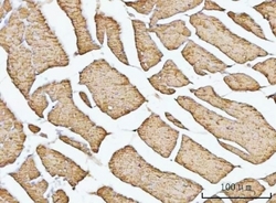
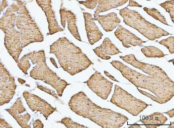
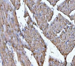
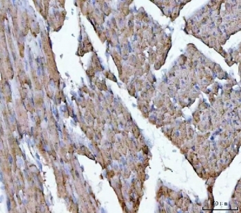
- IHC staining of FFPE mouse heart tissue with VDAC1 antibody. HIER: boil tissue sections in pH8 EDTA for 20 min and allow to cool before testing.
- Submitted by
- NSJ Bioreagents (provider)
- Main image
- Experimental details
- IHC staining of FFPE rat heart tissue with VDAC1 antibody. HIER: boil tissue sections in pH8 EDTA for 20 min and allow to cool before testing.
Supportive validation
- Submitted by
- NSJ Bioreagents (provider)
- Main image
- Experimental details
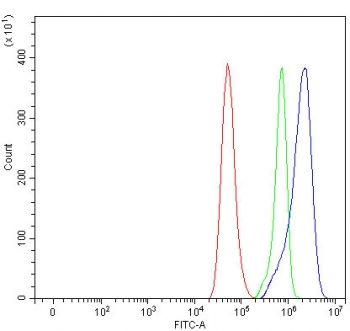
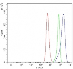
- Flow cytometry testing of fixed and permeabilized human HeLa cells with VDAC1 antibody at 1ug/million cells (blocked with goat sera); Red=cells alone, Green=isotype control, Blue= VDAC1 antibody.