 Explore
 Validate
 Learn
Learn Western blot
 Immunocytochemistry
 Immunoprecipitation
Antibody data
- Antibody Data
- Antigen structure
- References [1]
- Comments [0]
- Validations
- Immunocytochemistry [3]
- Immunohistochemistry [1]
Submit
Validation data
Reference
Comment
Report error
- Product number
- PA1-20988 - Provider product page

- Provider
- Invitrogen Antibodies
- Product name
- Anti-alpha Tubulin Polyclonal Antibody
- Antibody type
- Polyclonal
- Antigen
- Synthetic peptide
- Description
- PA1-20991 detects Wilm's tumor in Human, Mouse and Rat samples. PA1-20991 has been successfully used in Immunohistochemistry (Paraffin) procedures. Predicted molecular weight of 51 kDa. PA1-20991 immunogen corresponds to Synthetic peptide (Human) (C terminal).
- Reactivity
- Human, Mouse, Rat, Bovine, Canine, Guinea Pig, Porcine, Xenopus
- Host
- Rabbit
- Isotype
- IgG
- Vial size
- 500 µL
- Concentration
- 0.2 mg/ml
- Storage
- 4° C, do not freeze
Submitted references The DNA cytosine deaminase APOBEC3H haplotype I likely contributes to breast and lung cancer mutagenesis.
Starrett GJ, Luengas EM, McCann JL, Ebrahimi D, Temiz NA, Love RP, Feng Y, Adolph MB, Chelico L, Law EK, Carpenter MA, Harris RS
Nature communications 2016 Sep 21;7:12918
Nature communications 2016 Sep 21;7:12918
No comments: Submit comment
Supportive validation
- Submitted by
- Invitrogen Antibodies (provider)
- Main image
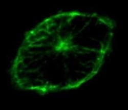
- Experimental details
- Immunofluorescent analysis of alpha Tubulin in z-section of a primary porcine macrophage using an Alpha Tubulin polyclonal antibody (Product # PA1-20988).

- Submitted by
- Invitrogen Antibodies (provider)
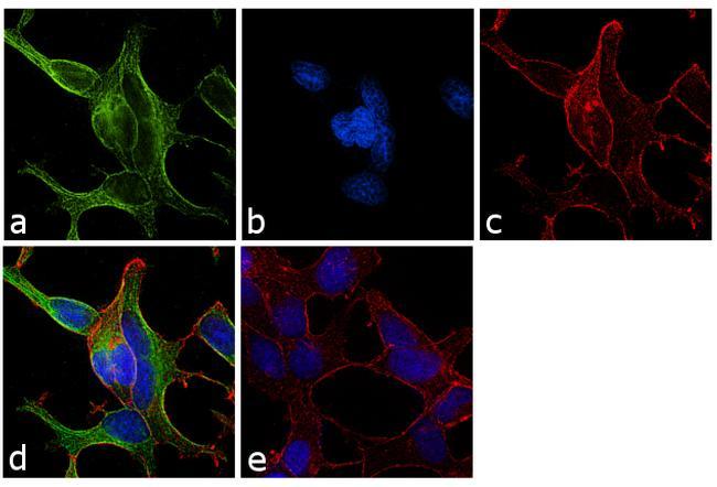
- Main image

- Experimental details
- Immunofluorescence analysis of alpha Tubulin was performed using 70% confluent log phase LNCaP cells. The cells were fixed with 4% paraformaldehyde for 10 minutes, permeabilized with 0.1% Triton™ X-100 for 10 minutes, and blocked with 1% BSA for 1 hour at room temperature. The cells were labeled with Alpha Tubulin Rabbit Polyclonal Antibody (Product # PA1-20988) at 2 µg/mL in 0.1% BSA and incubated for 3 hours at room temperature and then labeled with Goat anti-Rabbit IgG (H+L) Superclonal™ Secondary Antibody, Alexa Fluor® 488 conjugate (Product # A27034) at a dilution of 1:2000 for 45 minutes at room temperature (Panel a: green). Nuclei (Panel b: blue) were stained with SlowFade® Gold Antifade Mountant with DAPI (Product # S36938). F-actin (Panel c: red) was stained with Rhodamine Phalloidin (Product # R415, 1:300). Panel d represents the merged image showing cytoplasmic localization. Panel e shows the no primary antibody control. The images were captured at 60X magnification.
- Submitted by
- Invitrogen Antibodies (provider)
- Main image
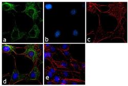
- Experimental details
- Immunofluorescence analysis of alpha Tubulin was performed using 70% confluent log phase NIH/3T3 cells. The cells were fixed with 4% paraformaldehyde for 10 minutes, permeabilized with 0.1% Triton™ X-100 for 10 minutes, and blocked with 1% BSA for 1 hour at room temperature. The cells were labeled with Alpha Tubulin Rabbit Polyclonal Antibody (Product # PA1-20988) at 2 µg/mL in 0.1% BSA and incubated for 3 hours at room temperature and then labeled with Goat anti-Rabbit IgG (H+L) Superclonal™ Secondary Antibody, Alexa Fluor® 488 conjugate (Product # A27034) at a dilution of 1:2000 for 45 minutes at room temperature (Panel a: green). Nuclei (Panel b: blue) were stained with SlowFade® Gold Antifade Mountant with DAPI (Product # S36938). F-actin (Panel c: red) was stained with Rhodamine Phalloidin (Product # R415, 1:300). Panel d represents the merged image showing cytoplasmic localization. Panel e shows the no primary antibody control. The images were captured at 60X magnification.

Supportive validation
- Submitted by
- Invitrogen Antibodies (provider)
- Main image
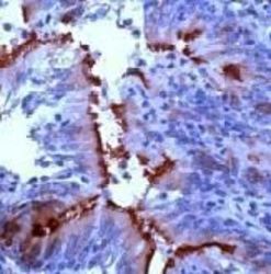
- Experimental details
- Immunohistochemical analysis of alpha Tubulin in human lung tissue using an Alpha Tubulin polyclonal antibody (Product # PA1-20988).
