Antibody data
- Antibody Data
- Antigen structure
- References [7]
- Comments [0]
- Validations
- Western blot [1]
- Immunoprecipitation [1]
- Immunohistochemistry [1]
Submit
Validation data
Reference
Comment
Report error
- Product number
- NB100-61071 - Provider product page

- Provider
- Novus Biologicals
- Proper citation
- Novus Cat#NB100-61071, RRID:AB_2283553
- Product name
- Rabbit Polyclonal Pericentrin Antibody
- Antibody type
- Polyclonal
- Description
- Immunogen affinity purified.
- Reactivity
- Human
- Host
- Rabbit
- Isotype
- IgG
- Vial size
- 100 ul
- Concentration
- 1.0 mg/ml
- Storage
- Store at 4C. Do not freeze.
Submitted references The C7orf43/TRAPPC14 component links the TRAPPII complex to Rabin8 for preciliary vesicle tethering at the mother centriole during ciliogenesis.
Akt Regulates a Rab11-Effector Switch Required for Ciliogenesis.
Investigation of F-BAR domain PACSIN proteins uncovers membrane tubulation function in cilia assembly and transport.
Neurymenolide A, a Novel Mitotic Spindle Poison from the New Caledonian Rhodophyta Phacelocarpus neurymenioides.
A LCMT1-PME-1 methylation equilibrium controls mitotic spindle size.
Reduced cilia frequencies in human renal cell carcinomas versus neighboring parenchymal tissue.
Neuropeptide Y family receptors traffic via the Bardet-Biedl syndrome pathway to signal in neuronal primary cilia.
Cuenca A, Insinna C, Zhao H, John P, Weiss MA, Lu Q, Walia V, Specht S, Manivannan S, Stauffer J, Peden AA, Westlake CJ
The Journal of biological chemistry 2019 Oct 18;294(42):15418-15434
The Journal of biological chemistry 2019 Oct 18;294(42):15418-15434
Akt Regulates a Rab11-Effector Switch Required for Ciliogenesis.
Walia V, Cuenca A, Vetter M, Insinna C, Perera S, Lu Q, Ritt DA, Semler E, Specht S, Stauffer J, Morrison DK, Lorentzen E, Westlake CJ
Developmental cell 2019 Jul 22;50(2):229-246.e7
Developmental cell 2019 Jul 22;50(2):229-246.e7
Investigation of F-BAR domain PACSIN proteins uncovers membrane tubulation function in cilia assembly and transport.
Insinna C, Lu Q, Teixeira I, Harned A, Semler EM, Stauffer J, Magidson V, Tiwari A, Kenworthy AK, Narayan K, Westlake CJ
Nature communications 2019 Jan 25;10(1):428
Nature communications 2019 Jan 25;10(1):428
Neurymenolide A, a Novel Mitotic Spindle Poison from the New Caledonian Rhodophyta Phacelocarpus neurymenioides.
Motuhi SE, Feizbakhsh O, Foll-Josselin B, Baratte B, Delehouzé C, Cousseau A, Fant X, Bulinski JC, Payri CE, Ruchaud S, Mehiri M, Bach S
Marine drugs 2019 Feb 1;17(2)
Marine drugs 2019 Feb 1;17(2)
A LCMT1-PME-1 methylation equilibrium controls mitotic spindle size.
Xia X, Gholkar A, Senese S, Torres JZ
Cell cycle (Georgetown, Tex.) 2015;14(12):1938-47
Cell cycle (Georgetown, Tex.) 2015;14(12):1938-47
Reduced cilia frequencies in human renal cell carcinomas versus neighboring parenchymal tissue.
Basten SG, Willekers S, Vermaat JS, Slaats GG, Voest EE, van Diest PJ, Giles RH
Cilia 2013 Jan 31;2(1):2
Cilia 2013 Jan 31;2(1):2
Neuropeptide Y family receptors traffic via the Bardet-Biedl syndrome pathway to signal in neuronal primary cilia.
Loktev AV, Jackson PK
Cell reports 2013 Dec 12;5(5):1316-29
Cell reports 2013 Dec 12;5(5):1316-29
No comments: Submit comment
Supportive validation
- Submitted by
- Novus Biologicals (provider)
- Main image

- Experimental details
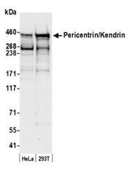
- Western Blot: Pericentrin Antibody [NB100-61071] - Detection of Human Pericentrin/Kendrin by Western Blot. Samples: Whole cell lysate (50 ug) from HeLa and 293T cells prepared using NETN lysis buffer. Antibody: Affinity purified rabbit anti-Pericentrin/Kendrin antibody NB100-61071 used for WB at 0.1 ug/ml. Detection: Chemiluminescence with an exposure time of 30 seconds.
Supportive validation
- Submitted by
- Novus Biologicals (provider)
- Main image

- Experimental details
- Immunoprecipitation: Pericentrin Antibody [NB100-61071] - Detection of human Pericentrin/Kendrin by western blot of immunoprecipitates. Samples: Whole cell lysate (0.5 or 1.0 mg per IP reaction; 20% of IP loaded) from HeLa cells prepared using NETN lysis buffer. Antibodies: Affinity purified rabbit anti-Pericentrin/Kendrin antibody NB100-61071 (lot 3) used for IP at 6 ug per reaction. Pericentrin/Kendrin was also immunoprecipitated by a previous lot of this antibody (lot 1). For blotting immunoprecipitated Pericentrin/Kendrin, NB100-61071 was used at 1 ug/ml. Detection: Chemiluminescence with an exposure time of 30 seconds.
Supportive validation
- Submitted by
- Novus Biologicals (provider)
- Main image

- Experimental details
- Immunohistochemistry: Pericentrin Antibody [NB100-61071] - Immunofluorescent analysis of cilia in renal tissues. Sections (4 um) of renal parenchymal tissues and tumor tissues were stained with DAPI, acetylated-alpha-tubulin (Ac-tub) and pericentrin (PCNT) to mark cell nuclei and cilia. Presented images are maximal projections of confocal images of typical parenchymal tissue and a representative ccRCC. Scale bars 20 um. Statistics were determined by performing a paired t-test at a 95% confidence interval. Image collected and cropped by CiteAb from the following publication (http://ciliajournal.biomedcentral.com/articles/10.1186/2046-2530-2-2) licensed under a CC-BY licence.
 Explore
 Validate
 Learn
Learn Western blot
 Immunocytochemistry