 Explore
 Validate
 Learn
Learn Western blot
Antibody data
- Antibody Data
- Antigen structure
- References [9]
- Comments [0]
- Validations
- Western blot [3]
- Immunohistochemistry [1]
Submit
Validation data
Reference
Comment
Report error
- Product number
- AF1034 - Provider product page

- Provider
- R&D Systems
- Product name
- Mouse/Rat Cathepsin C/DPPI Antibody
- Antibody type
- Polyclonal
- Description
- Antigen Affinity-purified. Detects mouse Cathepsin C/DPPI in direct ELISAs and Western blots. In direct ELISAs, approximately 15% cross-reactivity with recombinant human (rh) Cathepsin C is observed and less than 1% cross-reactivity with recombinant mouse (rm) Cathepsin A, rmCathepsin B, rmCathepsin D, rmCathepsin H, and rmCathepsin X/Z/P is observed.
- Reactivity
- Mouse, Rat
- Host
- Goat
- Conjugate
- Unconjugated
- Antigen sequence
P97821- Isotype
- IgG
- Vial size
- 100 ug
- Concentration
- LYOPH
- Storage
- Use a manual defrost freezer and avoid repeated freeze-thaw cycles. 12 months from date of receipt, -20 to -70 °C as supplied. 1 month, 2 to 8 °C under sterile conditions after reconstitution. 6 months, -20 to -70 °C under sterile conditions after reconstitution.
Submitted references The balance between cathepsin C and cystatin F controls remyelination in the brain of Plp1-overexpressing mouse, a chronic demyelinating disease model.
Deficiency for the cysteine protease cathepsin L impairs Myc-induced tumorigenesis in a mouse model of pancreatic neuroendocrine cancer.
Lysosomal protein turnover contributes to the acquisition of TGFβ-1 induced invasive properties of mammary cancer cells.
The endolysosomal cysteine cathepsins L and K are involved in macrophage-mediated clearance of Staphylococcus aureus and the concomitant cytokine induction.
Up-regulation of microglial cathepsin C expression and activity in lipopolysaccharide-induced neuroinflammation.
Gene targeting of the cysteine peptidase cathepsin H impairs lung surfactant in mice.
Mice deficient in LMAN1 exhibit FV and FVIII deficiencies and liver accumulation of α1-antitrypsin.
A role for serglycin proteoglycan in mast cell apoptosis induced by a secretory granule-mediated pathway.
Macrophages and cathepsin proteases blunt chemotherapeutic response in breast cancer.
Shimizu T, Wisessmith W, Li J, Abe M, Sakimura K, Chetsawang B, Sahara Y, Tohyama K, Tanaka KF, Ikenaka K
Glia 2017 Jun;65(6):917-930
Glia 2017 Jun;65(6):917-930
Deficiency for the cysteine protease cathepsin L impairs Myc-induced tumorigenesis in a mouse model of pancreatic neuroendocrine cancer.
Brindle NR, Joyce JA, Rostker F, Lawlor ER, Swigart-Brown L, Evan G, Hanahan D, Shchors K
PloS one 2015;10(4):e0120348
PloS one 2015;10(4):e0120348
Lysosomal protein turnover contributes to the acquisition of TGFβ-1 induced invasive properties of mammary cancer cells.
Kern U, Wischnewski V, Biniossek ML, Schilling O, Reinheckel T
Molecular cancer 2015 Feb 15;14:39
Molecular cancer 2015 Feb 15;14:39
The endolysosomal cysteine cathepsins L and K are involved in macrophage-mediated clearance of Staphylococcus aureus and the concomitant cytokine induction.
Müller S, Faulhaber A, Sieber C, Pfeifer D, Hochberg T, Gansz M, Deshmukh SD, Dauth S, Brix K, Saftig P, Peters C, Henneke P, Reinheckel T
FASEB journal : official publication of the Federation of American Societies for Experimental Biology 2014 Jan;28(1):162-75
FASEB journal : official publication of the Federation of American Societies for Experimental Biology 2014 Jan;28(1):162-75
Up-regulation of microglial cathepsin C expression and activity in lipopolysaccharide-induced neuroinflammation.
Fan K, Wu X, Fan B, Li N, Lin Y, Yao Y, Ma J
Journal of neuroinflammation 2012 May 20;9:96
Journal of neuroinflammation 2012 May 20;9:96
Gene targeting of the cysteine peptidase cathepsin H impairs lung surfactant in mice.
Bühling F, Kouadio M, Chwieralski CE, Kern U, Hohlfeld JM, Klemm N, Friedrichs N, Roth W, Deussing JM, Peters C, Reinheckel T
PloS one 2011;6(10):e26247
PloS one 2011;6(10):e26247
Mice deficient in LMAN1 exhibit FV and FVIII deficiencies and liver accumulation of α1-antitrypsin.
Zhang B, Zheng C, Zhu M, Tao J, Vasievich MP, Baines A, Kim J, Schekman R, Kaufman RJ, Ginsburg D
Blood 2011 Sep 22;118(12):3384-91
Blood 2011 Sep 22;118(12):3384-91
A role for serglycin proteoglycan in mast cell apoptosis induced by a secretory granule-mediated pathway.
Melo FR, Waern I, Rönnberg E, Åbrink M, Lee DM, Schlenner SM, Feyerabend TB, Rodewald HR, Turk B, Wernersson S, Pejler G
The Journal of biological chemistry 2011 Feb 18;286(7):5423-33
The Journal of biological chemistry 2011 Feb 18;286(7):5423-33
Macrophages and cathepsin proteases blunt chemotherapeutic response in breast cancer.
Shree T, Olson OC, Elie BT, Kester JC, Garfall AL, Simpson K, Bell-McGuinn KM, Zabor EC, Brogi E, Joyce JA
Genes & development 2011 Dec 1;25(23):2465-79
Genes & development 2011 Dec 1;25(23):2465-79
No comments: Submit comment
Supportive validation
- Submitted by
- R&D Systems (provider)
- Main image

- Experimental details
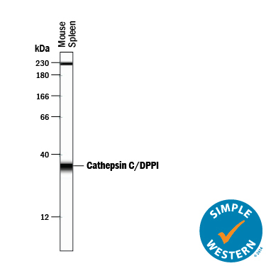
- Detection of Mouse Cathepsin C/DPPI by Simple WesternTM. Simple Western lane view shows lysates of mouse spleen tissue, loaded at 0.2 mg/mL. A specific band was detected for Cathepsin C/DPPI at approximately 35 kDa (as indicated) using 2.5 µg/mL of Goat Anti-Mouse Cathepsin C/DPPI Antigen Affinity-purified Polyclonal Antibody (Catalog # AF1034) followed by 1:50 dilution of HRP-conjugated Anti-Goat IgG Secondary Antibody (Catalog # HAF109). This experiment was conducted under reducing conditions and using the 12-230 kDa separation system. Non-specific interaction with the 230 kDa Simple Western standard may be seen with this antibody.
- Submitted by
- R&D Systems (provider)
- Main image
- Experimental details
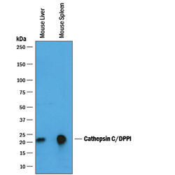
- Detection of Mouse Cathepsin C/DPPI by Western Blot. Western blot shows lysates of mouse liver tissue and mouse spleen tissue. PVDF membrane was probed with 0.25 µg/mL of Goat Anti-Mouse Cathepsin C/DPPI Antigen Affinity-purified Polyclonal Antibody (Catalog # AF1034) followed by HRP-conjugated Anti-Goat IgG Secondary Antibody (Catalog # HAF019). A specific band was detected for Cathepsin C/DPPI at approximately 20-25 kDa (as indicated). This experiment was conducted under reducing conditions and using Immunoblot Buffer Group 1.
- Submitted by
- R&D Systems (provider)
- Main image
- Experimental details
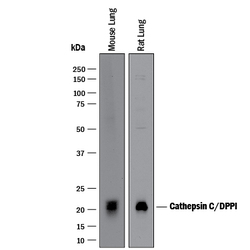
- Detection of Mouse and Rat Cathepsin C/DPPI by Western Blot. Western blot shows lysates of mouse lung tissue and rat lung tissue. PVDF membrane was probed with 1 µg/mL of Goat Anti-Mouse Cathepsin C/DPPI Antigen Affinity-purified Polyclonal Antibody (Catalog # AF1034) followed by HRP-conjugated Anti-Goat IgG Secondary Antibody (Catalog # HAF017). A specific band was detected for Cathepsin C/DPPI at approximately 20-22 kDa (as indicated). This experiment was conducted under reducing conditions and using Immunoblot Buffer Group 1.

Supportive validation
- Submitted by
- R&D Systems (provider)
- Main image
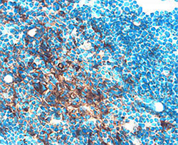
- Experimental details
- Cathepsin C/DPPI in Mouse Thymus. Cathepsin C/DPPI was detected in perfusion fixed frozen sections of mouse thymus using Goat Anti-Mouse Cathepsin C/ DPPI Antigen Affinity-purified Polyclonal Antibody (Catalog # AF1034) at 15 µg/mL overnight at 4 °C. Tissue was stained using the Anti-Goat HRP-DAB Cell & Tissue Staining Kit (brown; Catalog # CTS008) and counter-stained with hematoxylin (blue). Specific staining was localized to thymocytes. View our protocol for Chromogenic IHC Staining of Frozen Tissue Sections.
