 Explore
 Validate
 Learn
Learn Western blot
 ELISA
ELISA Immunocytochemistry
 Immunohistochemistry
Antibody data
- Antibody Data
- Antigen structure
- References [0]
- Comments [0]
- Validations
- Western blot [1]
- Immunohistochemistry [2]
Submit
Validation data
Reference
Comment
Report error
- Product number
- LS-B10591 - Provider product page

- Provider
- LSBio
- Product name
- IHC-plus™ TUBA1B / Tubulin Alpha 1B Antibody (C-Terminus, clone 17H11.F10) LS-B10591
- Antibody type
- Monoclonal
- Description
- Protein A purified
- Reactivity
- Human, Mouse, Rat, Bovine, Chicken/Avian
- Host
- Mouse
- Isotype
- IgG
- Antibody clone number
- 17H11.F10
- Storage
- Short term: store at 4°C. Long term: aliquot and store at -20°C. Avoid freeze-thaw cycles.
No comments: Submit comment
Supportive validation
- Submitted by
- LSBio (provider)
- Enhanced method
- Genetic validation
- Main image
- Experimental details
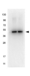
- Anti-alpha-Tubulin Monoclonal Antibody - Western Blot. HeLa whole cell lysate (left lane) and HEK293 whole cell lysate (right lane) were loaded with 10 ug of lysate each. The blot was blocked with Blocking Buffer (MB-070) for 30 min at RT, then washed and incubated with anti-Tubulin monoclonal antibody diluted in Blocking Buffer (p/n MB-070) at 1:1000 for 1 h at RT. After washing, blot was incubated with a 1:40000 dilution of HRP Rb a-Ms IgG (p/n LS-C60772) secondary antibody in Blocking Buffer (p/n MB-070) for 30 minutes at RT. Data was collected using Bio-Rad VersaDoc 4000 MP.

Enhanced validation
- Submitted by
- LSBio (provider)
- Enhanced method
- Genetic validation
- Main image

- Experimental details
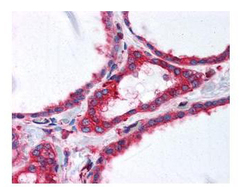
- Anti-alpha-Tubulin Monoclonal Antibody - Immunohistochemistry. anti-a-tubulin monoclonal antibody was used at a 2.5 ug/mL to detect tubulin in thyroid follicular epithelium (40X) showing moderate to strong cytoplasmic staining (image). Moderate to strong cytoplasmic staining was also observed within subsets of neurons and glia, and epithelial cells including adrenal, breast, colon, pancreas, kidney, prostate, placenta, skin, testis, uterus, thyroid, and within lymphoid organs. The image shows the localization of the antibody as the precipitated red signal, with a hematoxylin purple nuclear counterstain. tissue was formalin-fixed and paraffin embedded.

- Submitted by
- LSBio (provider)
- Enhanced method
- Genetic validation
- Main image
- Experimental details
- Anti-alpha-Tubulin Monoclonal Antibody - Immunohistochemistry. anti-a-tubulin monoclonal antibody was used at a 2.5 ug/mL to detect tubulin in thyroid follicular epithelium (40X) showing moderate to strong cytoplasmic staining (image). Moderate to strong cytoplasmic staining was also observed within subsets of neurons and glia, and epithelial cells including adrenal, breast, colon, pancreas, kidney, prostate, placenta, skin, testis, uterus, thyroid, and within lymphoid organs. The image shows the localization of the antibody as the precipitated red signal, with a hematoxylin purple nuclear counterstain. tissue was formalin-fixed and paraffin embedded.
