 Explore
 Validate
 Learn
Learn Western blot
Antibody data
- Antibody Data
- Antigen structure
- References [2]
- Comments [0]
- Validations
- Western blot [1]
- Immunocytochemistry [1]
- Immunohistochemistry [1]
Submit
Validation data
Reference
Comment
Report error
- Product number
- MAB8159 - Provider product page

- Provider
- Abnova Corporation
- Proper citation
- Abnova Corporation Cat#MAB8159, RRID:AB_10674385
- Product name
- TUBA1B monoclonal antibody, clone 17H11.F10
- Antibody type
- Monoclonal
- Description
- Mouse monoclonal antibody raised against synthetic peptide of TUBA1B.
- Antibody clone number
- 17H11.F10
- Storage
- Store at 4°C. For long term storage store at -20°C.Aliquot to avoid repeated freezing and thawing.
Submitted references Structural features and restricted expression of a human alpha-tubulin gene.
Expression of human alpha-tubulin genes: interspecies conservation of 3' untranslated regions.
Hall JL, Cowan NJ
Nucleic acids research 1985 Jan 11;13(1):207-23
Nucleic acids research 1985 Jan 11;13(1):207-23
Expression of human alpha-tubulin genes: interspecies conservation of 3' untranslated regions.
Cowan NJ, Dobner PR, Fuchs EV, Cleveland DW
Molecular and cellular biology 1983 Oct;3(10):1738-45
Molecular and cellular biology 1983 Oct;3(10):1738-45
No comments: Submit comment
Supportive validation
- Submitted by
- Abnova Corporation (provider)
- Main image
- Experimental details
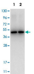
- Western Blotting from HeLa whole cell lysate (Lane 1) and HEK293 whole cell lysate (Lane 2) were loaded with 10 ug of lysate each. TUBA1B monoclonal antibody, clone 17H11.F10 (Cat # MAB8159) at 1 : 1,000 for 1 h at RT. Data was collected using Bio-Rad VersaDoc® 4000 MP.

Supportive validation
- Submitted by
- Abnova Corporation (provider)
- Main image

- Experimental details
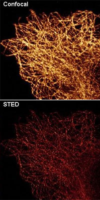
- Immunofluorescence staining of TUBA1B monoclonal antibody, clone 17H11.F10 (Cat # MAB8159) was used at a 0.1 ug/mL to detect TUBA1B in 4% paraformaldehyde fixed A-549 cells. Staining is shown using conventional confocal microscopy (upper panel) and by high resolution TCS STED nanoscopy (bottom panel). DyLight488™ conjugated anti-mouse IgG secondary antibody was used for detection at 1 ug/mL .Personal Communication, Myriam. Gastard, Leica Microsystems, Exton PA.
- Validation comment
- Immunofluorescence
Supportive validation
- Submitted by
- Abnova Corporation (provider)
- Main image
- Experimental details
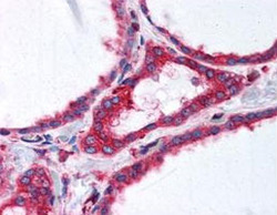
- Immunohistochemical staining of TUBA1B monoclonal antibody, clone 17H11.F10 (Cat # MAB8159) was used at a 2.5 ug/mL to detect TUBA1B in human thyroid follicular epithelium (40X) showing moderate to strong cytoplasmic staining (image). Moderate to strong cytoplasmic staining was also observed within subsets of neurons and glia, and epithelial cells including adrenal, breast, colon, pancreas, kidney, prostate, placenta, skin, testis, uterus, thyroid, and within lymphoid organs. The image shows the localization of the antibody as the precipitated red signal, with a hematoxylin purple nuclear counterstain. Tissue was formalin-fixed and paraffin embedded.Personal Communication, Vasiliki Demas, LifeSpanBiosciences, Seattle, WA.
- Validation comment
- Immunohistochemistry (Formalin/PFA-fixed paraffin-embedded sections)
