Explore
 Validate
 Learn
Learn Western blot
 Immunocytochemistry
Antibody data
- Antibody Data
- Antigen structure
- References [1]
- Comments [0]
- Validations
- Western blot [2]
- Flow cytometry [1]
Submit
Validation data
Reference
Comment
Report error
- Product number
- NBP2-02272 - Provider product page

- Provider
- Novus Biologicals
- Product name
- Mouse Monoclonal Hexokinase 2 Antibody
- Antibody type
- Monoclonal
- Description
- Affinity purified. Human Hexokinase-2 protein is 74% identical with Hexokinase-1 and our Hexokinase 2 antibody (clone 4C5) is not expected to crossreact with Hexokinase-1.
- Reactivity
- Human, Canine
- Host
- Mouse
- Isotype
- IgG
- Vial size
- 0.1 ml
- Concentration
- 1 mg/ml
- Storage
- Store at -20C. Avoid freeze-thaw cycles.
Submitted references Role of pyruvate kinase M2-mediated metabolic reprogramming during podocyte differentiation.
Yuan Q, Miao J, Yang Q, Fang L, Fang Y, Ding H, Zhou Y, Jiang L, Dai C, Zen K, Sun Q, Yang J
Cell death & disease 2020 May 11;11(5):355
Cell death & disease 2020 May 11;11(5):355
No comments: Submit comment
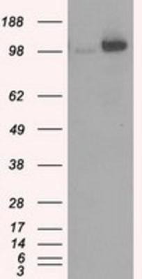
Supportive validation
- Submitted by
- Novus Biologicals (provider)
- Main image
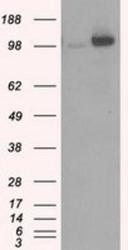
- Experimental details
- Western Blot: Hexokinase 2 Antibody (OTI4C5) [NBP2-02272] - HEK293T cells were transfected with the pCMV6-ENTRY control (Left lane) or pCMV6-ENTRY Hexokinase 2 (Right lane) cDNA for 48 hrs and lysed. Equivalent amounts of cell lysates (5 ug per lane) were separated by SDS-PAGE and immunoblotted with anti-Hexokinase 2.
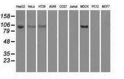
- Submitted by
- Novus Biologicals (provider)
- Main image

- Experimental details
- Western Blot: Hexokinase 2 Antibody (OTI4C5) [NBP2-02272] - Analysis of extracts (35ug) from 9 different cell lines by using anti-Hexokinase 2I monoclonal antibody.
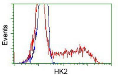
Supportive validation
- Submitted by
- Novus Biologicals (provider)
- Main image
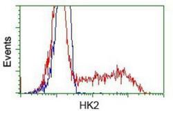
- Experimental details
- Flow Cytometry: Hexokinase 2 Antibody (OTI4C5) [NBP2-02272] - HEK293T cells transfected with either pCMV6-ENTRY Hexokinase 2.(Red) or empty vector control plasmid (Blue) were immunostained with anti-Hexokinase 2 mouse monoclonal, and then analyzed by flow cytometry.