Explore
 Validate
 Learn
Learn Western blot
Antibody data
- Antibody Data
- Antigen structure
- References [2]
- Comments [0]
- Validations
- Western blot [3]
- Immunocytochemistry [1]
- Immunohistochemistry [1]
- Other assay [2]
Submit
Validation data
Reference
Comment
Report error
- Product number
- PA5-31448 - Provider product page

- Provider
- Invitrogen Antibodies
- Product name
- PIWIL4 Polyclonal Antibody
- Antibody type
- Polyclonal
- Antigen
- Recombinant protein fragment
- Description
- Recommended positive controls: PIWIL4-transfected 293T. Predicted reactivity: Rat (80%), Pig (85%), Rhesus Monkey (98%). Store product as a concentrated solution. Centrifuge briefly prior to opening the vial.
- Reactivity
- Human
- Host
- Rabbit
- Isotype
- IgG
- Vial size
- 100 µL
- Concentration
- 0.69 mg/mL
- Storage
- Store at 4°C short term. For long term storage, store at -20°C, avoiding freeze/thaw cycles.
Submitted references Possible role of HIWI2 in modulating tight junction proteins in retinal pigment epithelial cells through Akt signaling pathway.
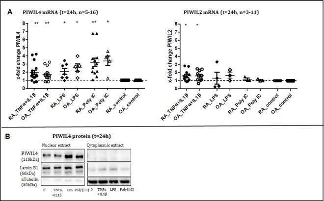
Expression and Regulation of PIWIL-Proteins and PIWI-Interacting RNAs in Rheumatoid Arthritis.
Sivagurunathan S, Palanisamy K, Arunachalam JP, Chidambaram S
Molecular and cellular biochemistry 2017 Mar;427(1-2):145-156
Molecular and cellular biochemistry 2017 Mar;427(1-2):145-156
Expression and Regulation of PIWIL-Proteins and PIWI-Interacting RNAs in Rheumatoid Arthritis.
Pleštilová L, Neidhart M, Russo G, Frank-Bertoncelj M, Ospelt C, Ciurea A, Kolling C, Gay RE, Michel BA, Vencovský J, Gay S, Jüngel A
PloS one 2016;11(11):e0166920
PloS one 2016;11(11):e0166920
No comments: Submit comment
Supportive validation
- Submitted by
- Invitrogen Antibodies (provider)
- Main image
- Experimental details
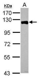
- Western blot analysis of PIWIL4 using 30 µg of MCF-7 lysate. Samples were loaded onto a 7.5% SDS-PAGE gel and probed with a PIWIL4 polyclonal antibody (Product # PA5-31448) at a dilution of 1:1000.

- Submitted by
- Invitrogen Antibodies (provider)
- Main image
- Experimental details
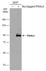
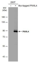
- Western Blot analysis of PIWIL4 was performed by separating 30 µg of non-transfected (–) and transfected (+) 293T whole cell extracts by 5% SDS-PAGE. Proteins were transferred to a membrane and probed with a PIWIL4 Polyclonal Antibody (Product # PA5-31448) at a dilution of 1:5000. The HRP-conjugated anti-rabbit IgG antibody was used to detect the primary antibody.
- Submitted by
- Invitrogen Antibodies (provider)
- Main image
- Experimental details
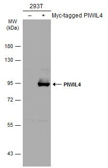
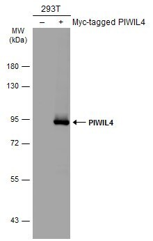
- Western Blot using PIWIL4 Polyclonal Antibody (Product # PA5-31448). Non-transfected (–) and transfected (+) 293T whole cell extracts (30 µg) were separated by 7.5% SDS-PAGE, and the membrane was blotted with PIWIL4 Polyclonal Antibody (Product # PA5-31448) diluted at 1:5,000. The HRP-conjugated anti-rabbit IgG antibody was used to detect the primary antibody.
Supportive validation
- Submitted by
- Invitrogen Antibodies (provider)
- Main image
- Experimental details
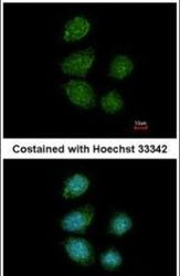
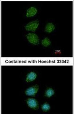
- Immunofluorescent analysis of PIWIL4 in paraformaldehyde-fixed MCF-7 cells using a PIWIL4 polyclonal antibody (Product # PA5-31448) at a 1:200 dilution.
Supportive validation
- Submitted by
- Invitrogen Antibodies (provider)
- Main image
- Experimental details
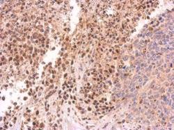
- Immunohistochemical analysis of paraffin-embedded 59T xenograft, using PIWIL4 (Product # PA5-31448) antibody at 1:500 dilution. Antigen Retrieval: EDTA based buffer, pH 8.0, 15 min.

Supportive validation
- Submitted by
- Invitrogen Antibodies (provider)
- Main image

- Experimental details
- NULL
- Submitted by
- Invitrogen Antibodies (provider)
- Main image
- Experimental details
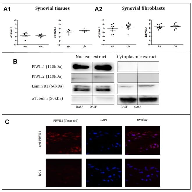
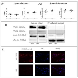
- Fig 1 Expression of PIWIL genes A1. mRNA expression of PIWIL2 and PIWIL4 in synovial tissues (ST) from RA and OA patients. Mean dCt PIWIL2 in RAST 5.3 and in OAST 5.5; mean dCt PIWIL4 in RAST 3.6 and in OAST 2.9. PIWIL1 and PIWIL3 were not expressed. A2. mRNA expression of PIWIL2 and PIWIL4 in isolated synovial fibroblasts (SF) from RA and OA patients. Mean dCt PIWIL2 in RASF 2.2 and in OASF 1.3; mean dCt PIWIL4 in RASF 1.3 and in OASF 1.2. PIWIL1 and PIWIL3 were not expressed. B. PIWIL4 protein was detected by Western blot in both RASF and OASF in the nucleus, but not in the cytoplasm. PIWIL2 expression was weak. C. Immunofluorescence has shown the presence of PIWIL4 protein predominantly in the cell nucleus and in the perinuclear regions (representative example of a RASF culture).