 Explore
 Validate
 Learn
Learn Western blot
Antibody data
- Antibody Data
- Antigen structure
- References [5]
- Comments [0]
- Validations
- Western blot [2]
- Immunocytochemistry [1]
- Immunohistochemistry [1]
Submit
Validation data
Reference
Comment
Report error
- Product number
- GTX119416 - Provider product page

- Provider
- GeneTex
- Proper citation
- GeneTex Cat#GTX119416, RRID:AB_10617835
- Product name
- VPS33A antibody [C1C3]
- Antibody type
- Polyclonal
- Reactivity
- Human, Mouse
- Host
- Rabbit
Submitted references Accumulation of undegraded autophagosomes by expression of dominant-negative STX17 (syntaxin 17) mutants.
LAMP-2 is required for incorporating syntaxin-17 into autophagosomes and for their fusion with lysosomes.
Characterization of the Mammalian CORVET and HOPS Complexes and Their Modular Restructuring for Endosome Specificity.
The HOPS complex mediates autophagosome-lysosome fusion through interaction with syntaxin 17.
Late endosomal transport and tethering are coupled processes controlled by RILP and the cholesterol sensor ORP1L.
Uematsu M, Nishimura T, Sakamaki Y, Yamamoto H, Mizushima N
Autophagy 2017 Aug 3;13(8):1452-1464
Autophagy 2017 Aug 3;13(8):1452-1464
LAMP-2 is required for incorporating syntaxin-17 into autophagosomes and for their fusion with lysosomes.
Hubert V, Peschel A, Langer B, Gröger M, Rees A, Kain R
Biology open 2016 Oct 15;5(10):1516-1529
Biology open 2016 Oct 15;5(10):1516-1529
Characterization of the Mammalian CORVET and HOPS Complexes and Their Modular Restructuring for Endosome Specificity.
van der Kant R, Jonker CT, Wijdeven RH, Bakker J, Janssen L, Klumperman J, Neefjes J
The Journal of biological chemistry 2015 Dec 18;290(51):30280-90
The Journal of biological chemistry 2015 Dec 18;290(51):30280-90
The HOPS complex mediates autophagosome-lysosome fusion through interaction with syntaxin 17.
Jiang P, Nishimura T, Sakamaki Y, Itakura E, Hatta T, Natsume T, Mizushima N
Molecular biology of the cell 2014 Apr;25(8):1327-37
Molecular biology of the cell 2014 Apr;25(8):1327-37
Late endosomal transport and tethering are coupled processes controlled by RILP and the cholesterol sensor ORP1L.
van der Kant R, Fish A, Janssen L, Janssen H, Krom S, Ho N, Brummelkamp T, Carette J, Rocha N, Neefjes J
Journal of cell science 2013 Aug 1;126(Pt 15):3462-74
Journal of cell science 2013 Aug 1;126(Pt 15):3462-74
No comments: Submit comment
Supportive validation
- Submitted by
- GeneTex (provider)
- Main image

- Experimental details
- Sample (30 ug of whole cell lysate) A: A549 7.5% SDS PAGE GTX119416 diluted at 1:1000
- Validation comment
- WB

- Submitted by
- GeneTex (provider)
- Main image

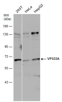
- Experimental details
- Various whole cell extracts (30 £gg) were separated by 7.5% SDS-PAGE, and the membrane was blotted with VPS33A antibody [C1C3] (GTX119416) diluted at 1:500. The signal was developed with Trident ECL plus-Enhanced.
Supportive validation
- Submitted by
- GeneTex (provider)
- Main image

- Experimental details
- VPS33A antibody [C1C3] detects VPS33A protein at cytoplasm by immunofluorescent analysis.Sample: HeLa cells were fixed in 4% paraformaldehyde at RT for 15 min.Green: VPS33A protein stained by VPS33A antibody [C1C3] (GTX119416) diluted at 1:200.Red: alpha Tubulin, a cytoskeleton marker, stained by alpha Tubulin antibody [GT114] (GTX628802) diluted at 1:1000.Blue: Hoechst 33342 staining.

Supportive validation
- Submitted by
- GeneTex (provider)
- Main image

- Experimental details
- Immunohistochemical analysis of paraffin-embedded human colon carcinoma, using VPS33A(GTX119416) antibody at 1:500 dilution.
