 Explore
 Validate
 Learn
Learn Western blot
 Immunoprecipitation
Antibody data
- Antibody Data
- Antigen structure
- References [0]
- Comments [0]
- Validations
- Western blot [1]
- Immunocytochemistry [1]
- Immunohistochemistry [2]
- Flow cytometry [1]
Submit
Validation data
Reference
Comment
Report error
- Product number
- PA5-72986 - Provider product page

- Provider
- Invitrogen Antibodies
- Product name
- SETX Polyclonal Antibody
- Antibody type
- Polyclonal
- Antigen
- Synthetic peptide
- Reactivity
- Human, Mouse
- Host
- Rabbit
- Isotype
- IgG
- Vial size
- 100 µL
- Concentration
- 1 mg/mL
- Storage
- Store at 4°C short term. For long term storage, store at -20°C, avoiding freeze/thaw cycles.
No comments: Submit comment
Supportive validation
- Submitted by
- Invitrogen Antibodies (provider)
- Main image
- Experimental details
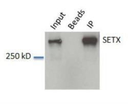
- Western blot analysis of SETX in HeLa whole cell lysate. Samples were incubated in SETX polyclonal antibody (Product # PA5-72986). Beads without antibody IP control. IP: IP from HeLa lysate.

Supportive validation
- Submitted by
- Invitrogen Antibodies (provider)
- Main image

- Experimental details
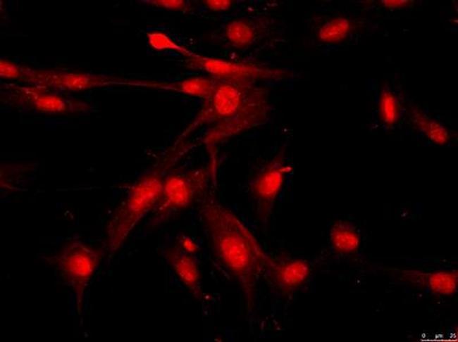
- Immunofluorescent analysis of fibroblasts cells using Senataxin polyclonal antibody (Product # PA5-72986).
Supportive validation
- Submitted by
- Invitrogen Antibodies (provider)
- Main image

- Experimental details
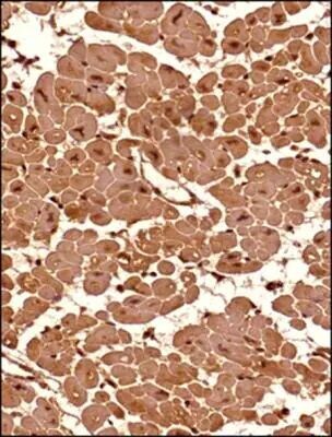
- Immunohistochemical analysis of SETX in formalin-fixed paraffin-embedded tissue section of human heart (transverse section). Samples were incubated in SETX polyclonal antibody (Product # PA5-72986) using a dilution of 1:200. The antibody generated a very strong staining in the cytoplasm and the nuclei of the muscle cells. No signal was found in the perimysium and endomycium area (connective tissue) of the section.
- Submitted by
- Invitrogen Antibodies (provider)
- Main image
- Experimental details
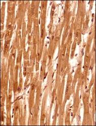
- Immunohistochemical analysis of SETX in formalin-fixed paraffin-embedded tissue section of human heart (verticle section). Samples were incubated in SETX polyclonal antibody (Product # PA5-72986) using a dilution of 1:200. The antibody generated a very strong staining in the cytoplasm and the nuclei of the muscle cells. No signal was found in the perimysium and endomycium (connective tissue) of the section.

Supportive validation
- Submitted by
- Invitrogen Antibodies (provider)
- Main image

- Experimental details
- Flow cytometry of SETX in Jurkat cells and a matched isotype control (orange). Samples were incubated in SETX polyclonal antibody (Product # PA5-72986) using a dilution of 2.5 µg/mL for 30 minutes at room temperature followed by a Rabbit IgG (H+L) Cross-Adsorbed secondary antibody. Cells were fixed with 4% PFA and then permeabilized with 0.1% saponin.
