 Explore
 Validate
 Learn
Learn Western blot
 Immunocytochemistry
Antibody data
- Antibody Data
- Antigen structure
- References [2]
- Comments [0]
- Validations
- Western blot [1]
- Immunohistochemistry [2]
- Flow cytometry [1]
Submit
Validation data
Reference
Comment
Report error
- Product number
- NBP1-94712 - Provider product page

- Provider
- Novus Biologicals
- Proper citation
- Novus Cat#NBP1-94712, RRID:AB_11010824
- Product name
- Rabbit Polyclonal Senataxin Antibody
- Antibody type
- Polyclonal
- Description
- Immunogen affinity purified.
- Reactivity
- Human, Mouse
- Host
- Rabbit
- Isotype
- IgG
- Vial size
- 0.1 ml
- Concentration
- 1.0 mg/ml
- Storage
- Store at 4C short term. Aliquot and store at -20C long term. Avoid freeze-thaw cycles.
Submitted references Altered translational repression of an RNA-binding protein, Elav by AOA2-causative Senataxin mutation.
A SUMO-dependent interaction between Senataxin and the exosome, disrupted in the neurodegenerative disease AOA2, targets the exosome to sites of transcription-induced DNA damage.
Choudhury SD, Vs A, Mushtaq Z, Kumar V
Synapse (New York, N.Y.) 2017 May;71(5)
Synapse (New York, N.Y.) 2017 May;71(5)
A SUMO-dependent interaction between Senataxin and the exosome, disrupted in the neurodegenerative disease AOA2, targets the exosome to sites of transcription-induced DNA damage.
Richard P, Feng S, Manley JL
Genes & development 2013 Oct 15;27(20):2227-32
Genes & development 2013 Oct 15;27(20):2227-32
No comments: Submit comment
Supportive validation
- Submitted by
- Novus Biologicals (provider)
- Main image
- Experimental details
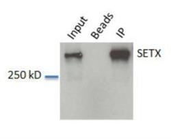
- Western Blot: Senataxin Antibody [NBP1-94712] - Input: HeLa whole cell lysate. Beads without antibody IP control. IP: IP from HeLa lysate.

Supportive validation
- Submitted by
- Novus Biologicals (provider)
- Main image
- Experimental details
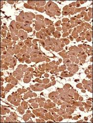
- Immunohistochemistry-Paraffin: Senataxin Antibody [NBP1-94712] - Analysis of a FFPE tissue section of human heart (transverse section) using 1:200 dilution of lot A2 of Senataxin antibody. The antibody generated a very strong staining in the cytoplasm and the nuclei of the muscle cells. No signal was found in the perimysium and endomycium area (connective tissue) of the section.
- Submitted by
- Novus Biologicals (provider)
- Main image
- Experimental details
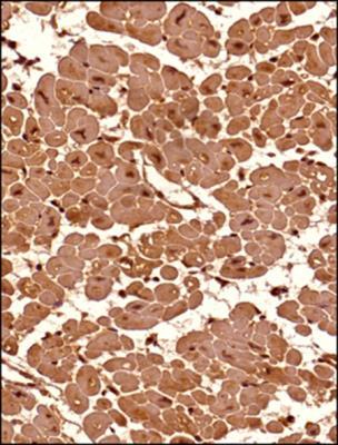
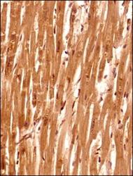
- Immunohistochemistry-Paraffin: Senataxin Antibody [NBP1-94712] - Analysis of a FFPE tissue section of human heart (verticle section) using 1:200 dilution of lot A2 of Senataxin antibody. The antibody generated a very strong staining in the cytoplasm and the nuclei of the muscle cells. No signal was found in the perimysium and endomycium (connective tissue) of the section.

Supportive validation
- Submitted by
- Novus Biologicals (provider)
- Main image

- Experimental details
- Flow Cytometry: Senataxin Antibody [NBP1-94712] - An intracellular stain was performed on Jurkat cells and a matched isotype control (orange). Cells were fixed with 4% PFA and then permeabilized with 0.1% saponin. Cells were incubated in an antibody dilution of 2.5 ug/mL for 30 minutes at room temperature, followed by Rabbit IgG (H+L) Cross-Adsorbed Secondary Antibody, .
