Explore
 Validate
 Learn
Learn Western blot
Antibody data
- Antibody Data
- Antigen structure
- References [1]
- Comments [0]
- Validations
- Western blot [2]
- Immunohistochemistry [6]
- Other assay [1]
Submit
Validation data
Reference
Comment
Report error
- Product number
- PA5-80195 - Provider product page

- Provider
- Invitrogen Antibodies
- Product name
- TSPAN12 Polyclonal Antibody
- Antibody type
- Polyclonal
- Antigen
- Recombinant full-length protein
- Description
- Reconstitute with 0.2 mL of distilled water to yield a concentration of 500 µg/mL.
- Reactivity
- Human, Mouse, Rat
- Host
- Rabbit
- Isotype
- IgG
- Vial size
- 100 µg
- Concentration
- 500 µg/mL
- Storage
- -20°C
Submitted references Ring finger protein 152-dependent degradation of TSPAN12 suppresses hepatocellular carcinoma progression.
Wan J, Liu S, Sun W, Yu H, Tang W, Liu W, Ji J, Liu B
Cancer cell international 2021 Feb 18;21(1):122
Cancer cell international 2021 Feb 18;21(1):122
No comments: Submit comment
Supportive validation
- Submitted by
- Invitrogen Antibodies (provider)
- Main image
- Experimental details
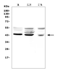
- Western blot analysis of TSPAN12 in, Lane 1: rat kidney tissue lysates, Lane 2: rat brain tissue lysates, Lane 3: mouse brain tissue lysates. Electrophoresis was performed on a 5-20% SDS-PAGE gel at 70V (Stacking gel) / 90V (Resolving gel) for 2-3 hours. The sample well of each lane was loaded with 50 µg of sample under reducing conditions. After Electrophoresis, proteins were transferred to a Nitrocellulose membrane at 150mA for 50-90 minutes. The membrane was blocked with 5% Non-fat Milk/ TBS for 1. 5 hour at RT. The membrane was incubated with TSPAN12 Polyclonal Antibody (Product # PA5-80195) at 0.5 μg/mL overnight at 4°C, then washed with TBS-0. 1% Tween 3 times with 5 minutes each and probed with a goat anti-rabbit IgG-HRP secondary antibody at a dilution of 1:10000 for 1. 5 hour at RT. The signal is developed using an Enhanced Chemiluminescent detection (ECL) kit. A specific band was detected for TSPAN12 at approximately 43KD. The expected band size for TSPAN12 is at 35KD.

- Submitted by
- Invitrogen Antibodies (provider)
- Main image
- Experimental details
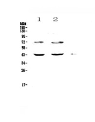
- Western blot analysis of TSPAN12 in Lane 1: human HeLa cell lysate, Lane 2: human SW620 cell lysate using 50 µg (reducing conditions) per well. Electrophoresis was performed on 5-20% SDS-PAGE gel at 70V (Stacking gel) / 90V (Resolving gel) for 2-3 hours and protein was transferred to a nitrocellulose membrane at 150mA for 50-90 minutes. Sample was blocked with 5% Non-fat Milk/TBS for 1.5 hours at room temperature, incubated with TSPAN12 polyclonal antibody (Product # PA5-80195) at a dilution of 0.5 µg/mL (overnight at 4°C), followed by goat anti-rabbit IgG-HRP secondary antibody at a dilution of 1:10,000. Signal development was performed using a chemiluminescence (ECL) kit.

Supportive validation
- Submitted by
- Invitrogen Antibodies (provider)
- Main image
- Experimental details
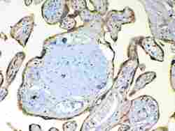
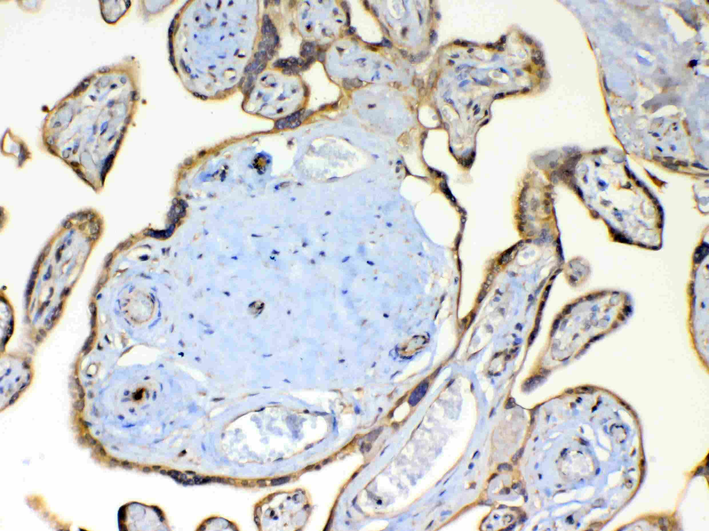
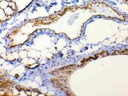
- Immunohistochemistry analysis of TSPAN12 on paraffin-embedded human placenta tissue. Antigen retrieval was performed using citrate buffer (pH6, epitope retrieval solution) for 20 mins. Sample was blocked using 10% goat serum, incubated with TSPAN12 polyclonal antibody (Product# PA5-80195) with a dilution of 1 µg/mL (overnight at 4°C), and followed by biotinylated goat anti-rabbit IgG (30 minutes at 37°C). Development was performed using Streptavidin-Biotin-Complex (SABC) with DAB chromogen method.
- Submitted by
- Invitrogen Antibodies (provider)
- Main image
- Experimental details
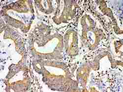
- Immunohistochemistry analysis of TSPAN12 on paraffin-embedded human rectal cancer tissue. Antigen retrieval was performed using citrate buffer (pH6, epitope retrieval solution) for 20 mins. Sample was blocked using 10% goat serum, incubated with TSPAN12 polyclonal antibody (Product# PA5-80195) with a dilution of 1 µg/mL (overnight at 4°C), and followed by biotinylated goat anti-rabbit IgG (30 minutes at 37°C). Development was performed using Streptavidin-Biotin-Complex (SABC) with DAB chromogen method.
- Submitted by
- Invitrogen Antibodies (provider)
- Main image
- Experimental details
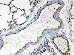
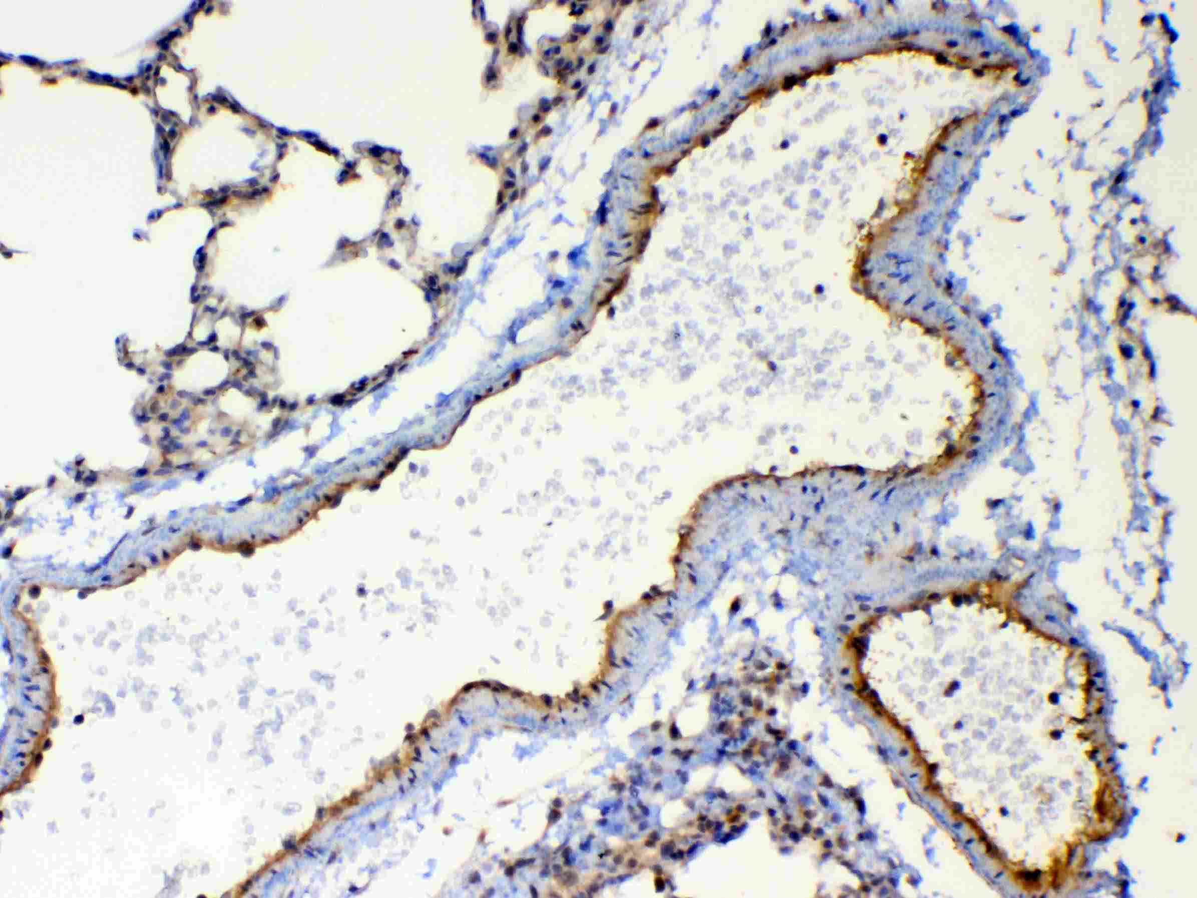
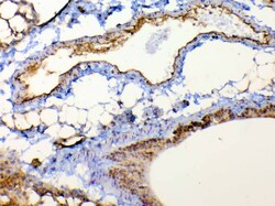
- Immunohistochemistry analysis of TSPAN12 on paraffin-embedded rat lung tissue. Antigen retrieval was performed using citrate buffer (pH6, epitope retrieval solution) for 20 mins. Sample was blocked using 10% goat serum, incubated with TSPAN12 polyclonal antibody (Product# PA5-80195) with a dilution of 1 µg/mL (overnight at 4°C), and followed by biotinylated goat anti-rabbit IgG (30 minutes at 37°C). Development was performed using Streptavidin-Biotin-Complex (SABC) with DAB chromogen method.
- Submitted by
- Invitrogen Antibodies (provider)
- Main image

- Experimental details
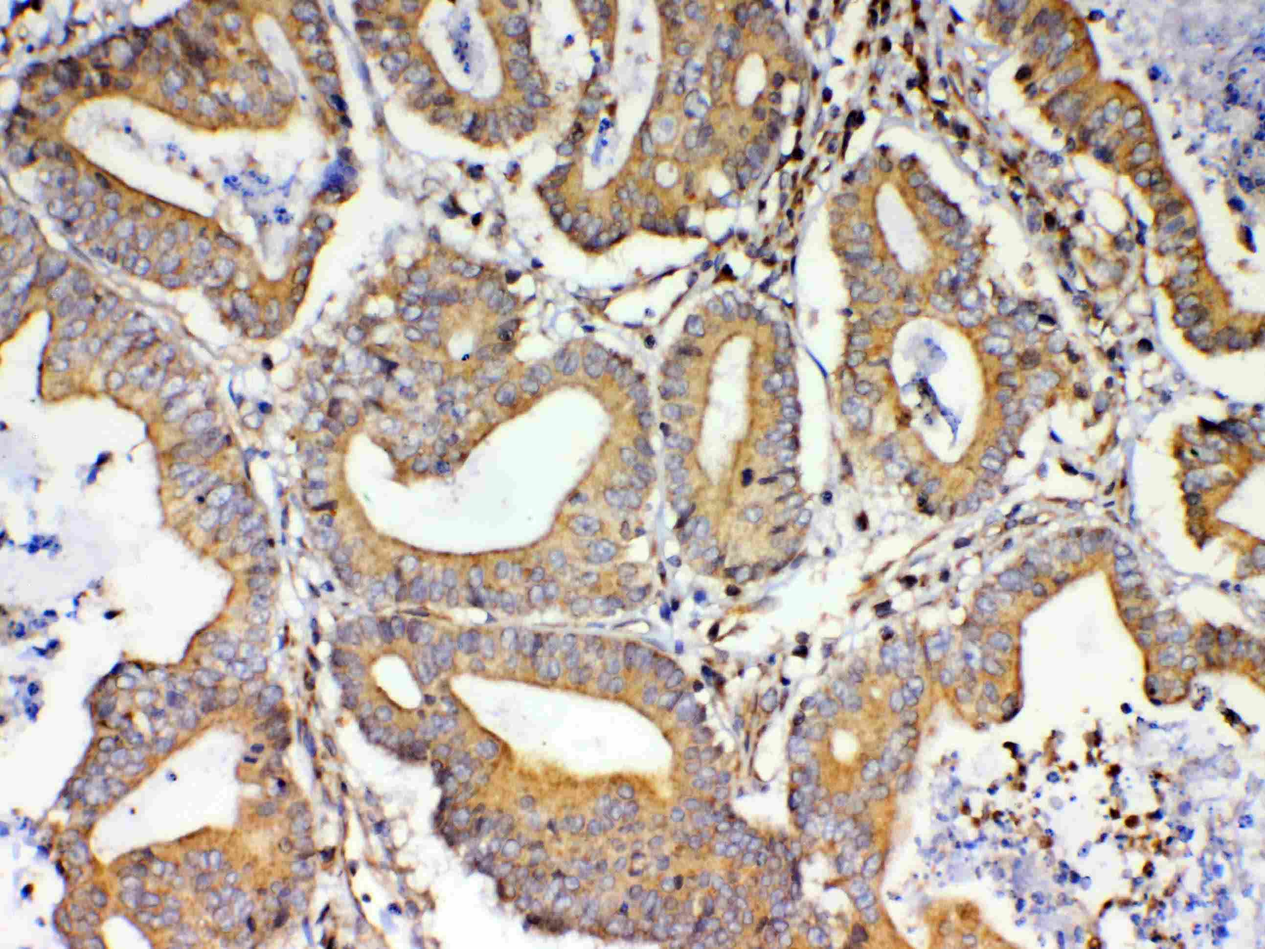
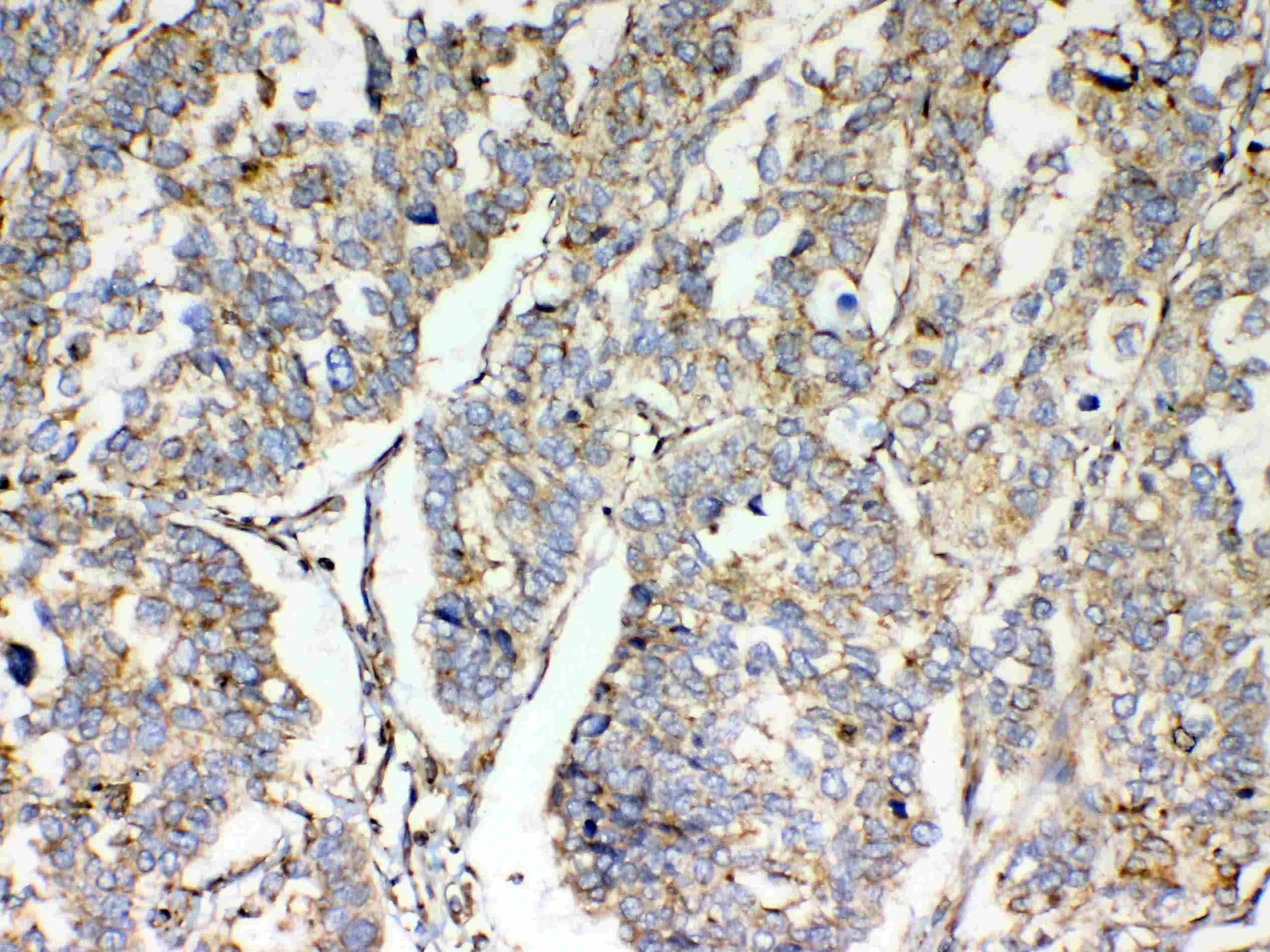
- Immunohistochemistry analysis of TSPAN12 on paraffin-embedded human lung cancer tissue. Antigen retrieval was performed using citrate buffer (pH6, epitope retrieval solution) for 20 mins. Sample was blocked using 10% goat serum, incubated with TSPAN12 polyclonal antibody (Product# PA5-80195) with a dilution of 1 µg/mL (overnight at 4°C), and followed by biotinylated goat anti-rabbit IgG (30 minutes at 37°C). Development was performed using Streptavidin-Biotin-Complex (SABC) with DAB chromogen method.
- Submitted by
- Invitrogen Antibodies (provider)
- Main image
- Experimental details
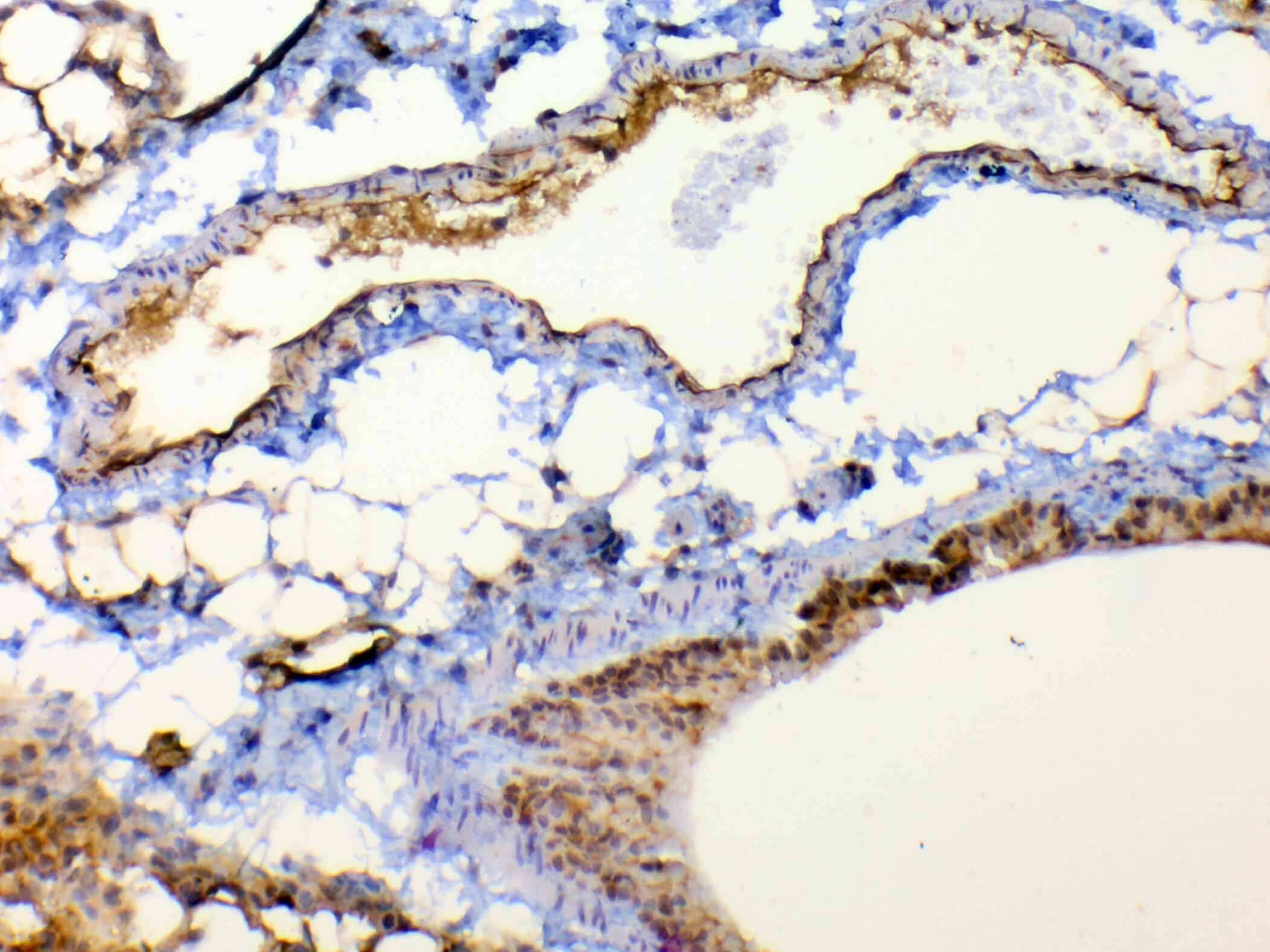
- Immunohistochemistry analysis of TSPAN12 on paraffin-embedded mouse lung tissue. Antigen retrieval was performed using citrate buffer (pH6, epitope retrieval solution) for 20 mins. Sample was blocked using 10% goat serum, incubated with TSPAN12 polyclonal antibody (Product# PA5-80195) with a dilution of 1 µg/mL (overnight at 4°C), and followed by biotinylated goat anti-rabbit IgG (30 minutes at 37°C). Development was performed using Streptavidin-Biotin-Complex (SABC) with DAB chromogen method.
- Submitted by
- Invitrogen Antibodies (provider)
- Main image
- Experimental details
- Immunohistochemistry analysis of TSPAN12 on paraffin-embedded mouse lung tissue. Antigen retrieval was performed using citrate buffer (pH6, epitope retrieval solution) for 20 mins. Sample was blocked using 10% goat serum, incubated with TSPAN12 polyclonal antibody (Product# PA5-80195) with a dilution of 1 µg/mL (overnight at 4°C), and followed by biotinylated goat anti-rabbit IgG (30 minutes at 37°C). Development was performed using Streptavidin-Biotin-Complex (SABC) with DAB chromogen method.

Supportive validation
- Submitted by
- Invitrogen Antibodies (provider)
- Main image

- Experimental details
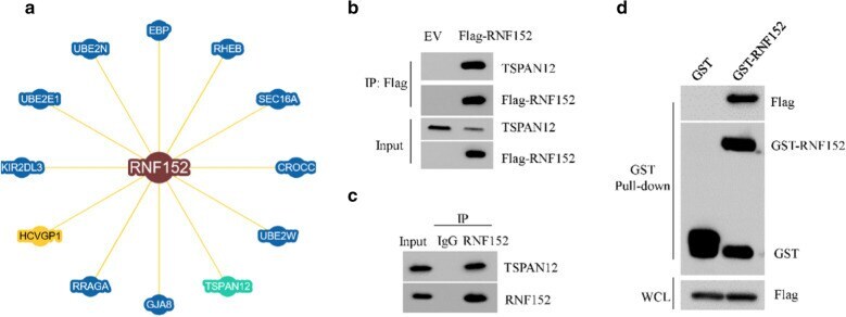
- Fig. 4 TSPAN12 was associated with RNF152. a The interaction protein network of RNF152 revealed by the BioGRID database ( https://thebiogrid.org/ ). b The cell lysates from 293 T cells transfected with empty vector (EV) or Flag-RNF152 were subjected to immunoprecipitation (IP) with Flag M2 beads, followed by immunoblotting with indicated antibodies. c The cell lysates of MG132-treated HuH6 cells were subjected to immunoprecipitation with IgG or anti-RNF152 antibody and detected by immunoblotting with indicated antibodies. d 293 T cells transfected Flag-tagged TSPAN12 plasmid were lysed and incubated with GST alone or GST-RNF152 immobilized on GST-Sepharose beads. The bound proteins were then detected by immunoblotting with indicated antibodies