 Explore
 Validate
 Learn
Learn Western blot
Antibody data
- Antibody Data
- Antigen structure
- References [1]
- Comments [0]
- Validations
- Western blot [1]
- Immunoprecipitation [1]
Submit
Validation data
Reference
Comment
Report error
- Product number
- NBP1-50042 - Provider product page

- Provider
- Novus Biologicals
- Proper citation
- Novus Cat#NBP1-50042, RRID:AB_10012232
- Product name
- Rabbit Polyclonal DENR Antibody
- Antibody type
- Polyclonal
- Description
- Immunogen affinity purified.
- Reactivity
- Human, Mouse
- Host
- Rabbit
- Isotype
- IgG
- Vial size
- 100 ul
- Concentration
- 1.0 mg/ml
- Storage
- Store at 4C. Do not freeze.
Submitted references De Novo Mutations in DENR Disrupt Neuronal Development and Link Congenital Neurological Disorders to Faulty mRNA Translation Re-initiation.
Haas MA, Ngo L, Li SS, Schleich S, Qu Z, Vanyai HK, Cullen HD, Cardona-Alberich A, Gladwyn-Ng IE, Pagnamenta AT, Taylor JC, Stewart H, Kini U, Duncan KE, Teleman AA, Keays DA, Heng JI
Cell reports 2016 Jun 7;15(10):2251-2265
Cell reports 2016 Jun 7;15(10):2251-2265
No comments: Submit comment
Supportive validation
- Submitted by
- Novus Biologicals (provider)
- Main image

- Experimental details
- Western Blot: DENR Antibody [NBP1-50042] - Detection of human and mouse DRP by western blot (h&m) and immunoprecipitation (h). Samples: Whole cell lysate from HeLa (5, 15 and 50 ug for WB; 1 mg for IP, 20% of IP loaded), HEK293T (T; 50 ug) and mouse NIH 3T3 (M; 50 ug) cells. Antibodies: Affinity purified rabbit anti-DRP antibody NBP1-50042 used for WB at 0.1 ug/ml (A) and 1 ug/ml (B) and used for IP at 6 ug/mg lysate. DRP was successfully immunoprecipitated by another rabbit anti-DRP antibody, which recognizes an upstream epitope. Detection: Chemiluminescence with exposure times of 30 seconds (A) and 10 seconds (B).

Supportive validation
- Submitted by
- Novus Biologicals (provider)
- Main image
- Experimental details
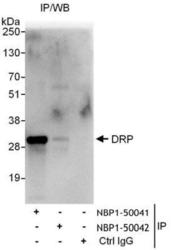
- Immunoprecipitation: DENR Antibody [NBP1-50042] - Samples: Whole cell lysate (1 mg for IP, 20% of IP loaded) from HeLa cells. Antibodies: Affinity purified rabbit anti-DRP antibody NBP1-50041 used for IP at 6 ug/mg lysate. DRP was not efficiently immunoprecipitated by rabbit anti-DRP antibody NBP1-50042, which recognizes a downstream epitope. For blotting immunoprecipitated DRP, NBP1-50042 was used at 1 ug/ml. Detection: Chemiluminescence with an exposure time of 10 seconds.
