 Explore
 Validate
 Learn
Learn Western blot
Antibody data
- Antibody Data
- Antigen structure
- References [0]
- Comments [0]
- Validations
- Western blot [5]
- Immunocytochemistry [2]
- Immunohistochemistry [1]
Submit
Validation data
Reference
Comment
Report error
- Product number
- PA5-22137 - Provider product page

- Provider
- Invitrogen Antibodies
- Product name
- ZC3H12A Polyclonal Antibody
- Antibody type
- Polyclonal
- Antigen
- Recombinant protein fragment
- Description
- Recommended positive controls: 293T, A431.
- Concentration
- 1.8 mg/mL
No comments: Submit comment
Supportive validation
- Submitted by
- Invitrogen Antibodies (provider)
- Main image

- Experimental details
- Western blot analysis of ZC3H12A using Various whole cell extracts (30 µg). Samples were loaded onto a 7.5% SDS-PAGE gel and probed with a ZC3H12A polyclonal antibody (Product # PA5-22137) at a dilution of 1:1000.

- Submitted by
- Invitrogen Antibodies (provider)
- Main image

- Experimental details
- Western Blot analysis of ZC3H12A was performed by separating 30 µg of various whole cell extracts by 7.5% SDS-PAGE. Proteins were transferred to a membrane and probed with a ZC3H12A Polyclonal Antibody (Product # PA5-22137) at a dilution of 1:1000 and a HRP-conjugated anti-rabbit IgG secondary antibody.

- Submitted by
- Invitrogen Antibodies (provider)
- Main image

- Experimental details
- Western blot was performed using Anti-ZC3H12A Polyclonal Antibody (Product # PA5-22137) and a ~65 kDa band corresponding to Endoribonuclease ZC3H12A was observed across cell lines and tissues tested . Whole cell extracts (30 µg lysate) of THP-1 (Lane 1), THP-1 treated with 1 µg/mL LPS for 16h (Lane 2), SK-O-V3 (Lane 3), Raji (Lane 4), SH-SY5Y (Lane 5), IMR-32 (Lane 6), Mouse Heart (Lane 7), were electrophoresed using NuPAGE™ 4-12% Bis-Tris Protein Gel (Product # NP0321BOX). Resolved proteins were then transferred onto a nitrocellulose membrane (Product # IB23001) by iBlot® 2 Dry Blotting System (Product # IB21001). The blot was probed with the primary antibody (1:1000) and detected by chemiluminescence with Goat anti-Rabbit IgG (H+L) Superclonal™ Recombinant Secondary Antibody, HRP (Product # A27036,1:20000 using the iBright™ FL1500 Imaging System (Product # A44115). Chemiluminescent detection was performed using SuperSignal™ West Pico PLUS Chemiluminescent Substrate (Product # 34580).Signal was induced in THP-1 cells treated with LPS as seen in lanes 1 and 2.

- Submitted by
- Invitrogen Antibodies (provider)
- Main image

- Experimental details
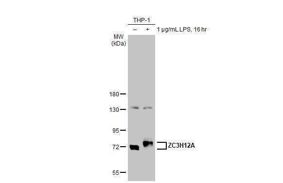
- Western Blot using ZC3H12A Polyclonal Antibody (Product # PA5-22137). Untreated (–) and treated (+) THP-1 whole cell extracts (30 µg) were separated by 7.5% SDS-PAGE, and the membrane was blotted with ZC3H12A Polyclonal Antibody (Product # PA5-22137) diluted at 1:1,000. The HRP-conjugated anti-rabbit IgG antibody was used to detect the primary antibody, and the signal was developed with Trident ECL plus-Enhanced.
- Submitted by
- Invitrogen Antibodies (provider)
- Main image

- Experimental details
- Knockdown of Endoribonuclease ZC3H12A was achieved by transfecting Raji with Endoribonuclease ZC3H12A specific siRNAs (Silencer® select Product # s36970, s36971). Western blot analysis (Fig. a) was performed using Whole cell extracts from the Endoribonuclease ZC3H12A knockdown cells (lane 3), non-targeting scrambled siRNA transfected cells (lane 2) and untransfected cells (lane 1). The blot was probed with ZC3H12A Polyclonal Antibody (Product # PA5-22137, 1:1000 ) and Goat anti-Rabbit IgG (H+L) Superclonal™ Recombinant Secondary Antibody, HRP (Product # A27036, 1:20000). Densitometric analysis of this western blot is shown in histogram (Fig. b). Decrease in signal upon siRNA mediated knock down confirms that antibody is specific to Endoribonuclease ZC3H12A.

Supportive validation
- Submitted by
- Invitrogen Antibodies (provider)
- Main image

- Experimental details
- Immunofluorescent analysis of ZC3H12A in paraformaldehyde-fixed HeLa cells using a ZC3H12A polyclonal antibody (Product # PA5-22137) at a 1:200 dilution.

- Submitted by
- Invitrogen Antibodies (provider)
- Main image

- Experimental details
- ZC3H12A antibody detects ZC3H12A protein at cytoplasm by immunofluorescent analysis. Sample: HeLa cells were fixed in 4% paraformaldehyde at RT for 15 min. Green: ZC3H12A stained by ZC3H12A antibody (Product # PA5-22137) diluted at 1:500.

Supportive validation
- Submitted by
- Invitrogen Antibodies (provider)
- Main image

- Experimental details
- Immunohistochemistry (Paraffin) analysis of ZC3H12A was performed in paraffin-embedded mouse kidney tissue using ZC3H12A Polyclonal Antibody (Product # PA5-22137) at a dilution of 1:250.
