 Explore
 Validate
 Learn
Learn Western blot
Antibody data
- Antibody Data
- Antigen structure
- References [3]
- Comments [0]
- Validations
- Western blot [3]
- Immunocytochemistry [1]
- Immunohistochemistry [7]
Submit
Validation data
Reference
Comment
Report error
- Product number
- TA500641 - Provider product page

- Provider
- OriGene
- Proper citation
- OriGene Cat#TA500641, RRID:AB_2168078
- Product name
- Anti-PRKAR1A (PKA regulatory subunit I alpha) mouse monoclonal antibody, clone OTI6C7 (formerly 6C7)
- Antibody type
- Monoclonal
- Description
- Anti-PRKAR1A (PKA regulatory subunit I alpha) mouse monoclonal antibody, clone OTI6C7 (formerly 6C7)
- Host
- Mouse
- Conjugate
- Unconjugated
- Epitope
- PRKAR1A
- Isotype
- IgG
- Antibody clone number
- OTI6C7
- Vial size
- 100 µl
- Concentration
- 1.00mg/ml
Submitted references Hepatic adenomas with synchronous or metachronous fibrolamellar carcinomas: both are characterized by LFABP loss.
Defects of the Carney complex gene (PRKAR1A) in odontogenic tumors.
PRKAR1A in the development of cardiac myxoma: a study of 110 cases including isolated and syndromic tumors.
Graham RP, Terracciano LM, Meves A, Vanderboom PM, Dasari S, Yeh MM, Torbenson MS, Cruise MW
Modern pathology : an official journal of the United States and Canadian Academy of Pathology, Inc 2016 Jun;29(6):607-15
Modern pathology : an official journal of the United States and Canadian Academy of Pathology, Inc 2016 Jun;29(6):607-15
Defects of the Carney complex gene (PRKAR1A) in odontogenic tumors.
Sousa SF, Gomez RS, Diniz MG, Bernardes VF, Soares FF, Brito JA, Liu S, Pontes HA, Stratakis CA, Gomes CC
Endocrine-related cancer 2015 Jun;22(3):399-408
Endocrine-related cancer 2015 Jun;22(3):399-408
PRKAR1A in the development of cardiac myxoma: a study of 110 cases including isolated and syndromic tumors.
Maleszewski JJ, Larsen BT, Kip NS, Castonguay MC, Edwards WD, Carney JA, Kipp BR
The American journal of surgical pathology 2014 Aug;38(8):1079-87
The American journal of surgical pathology 2014 Aug;38(8):1079-87
No comments: Submit comment
Supportive validation
- Submitted by
- OriGene (provider)
- Main image

- Experimental details
- Western blot analysis of extracts (35ug) from 9 different cell lines by using anti-PRKAR1A monoclonal antibody.
- Validation comment
- WB

- Submitted by
- OriGene (provider)
- Main image

- Experimental details
- HEK293T cells were transfected with the pCMV6-ENTRY control (Left lane) or pCMV6-ENTRY PRKAR1A (RC212810, Right lane) cDNA for 48 hrs and lysed. Equivalent amounts of cell lysates (5 ug per lane) were separated by SDS-PAGE and immunoblotted with anti-PRKAR1A.
- Validation comment
- WB

- Submitted by
- OriGene (provider)
- Main image

- Experimental details
- Western blot analysis of extracts (10ug) from a mouse cell line and 3 different mouse tissues by using anti-PRKAR1A monoclonal antibody.(1:200)
- Validation comment
- WB

Supportive validation
- Submitted by
- OriGene (provider)
- Main image

- Experimental details
- Anti-PRKAR1A mouse monoclonal antibody (TA500641) immunofluorescent staining of COS7 cells transiently transfected by pCMV6-ENTRY PRKAR1A(RC212810).
- Validation comment
- IF

Supportive validation
- Submitted by
- OriGene (provider)
- Main image

- Experimental details
- Immunohistochemical staining of paraffin-embedded endometrium tissue within the normal limits using anti-PRKAR1Amouse monoclonal antibody. (Heat-induced epitope retrieval by 10mM citric buffer, pH6.0, 100C for 10min, TA500641, Dilution 1:50)
- Validation comment
- IHC

- Submitted by
- OriGene (provider)
- Main image

- Experimental details
- Immunohistochemical staining of paraffin-embedded prostate tissue within the normal limits using anti-PRKAR1Amouse monoclonal antibody. (Heat-induced epitope retrieval by 10mM citric buffer, pH6.0, 100C for 10min, TA500641, Dilution 1:50)
- Validation comment
- IHC

- Submitted by
- OriGene (provider)
- Main image

- Experimental details
- Immunohistochemical staining of paraffin-embedded Carcinoma of bladder tissue using anti-PRKAR1Amouse monoclonal antibody. (Heat-induced epitope retrieval by 10mM citric buffer, pH6.0, 100C for 10min, TA500641, Dilution 1:50)
- Validation comment
- IHC

- Submitted by
- OriGene (provider)
- Main image

- Experimental details
- Immunohistochemical staining of paraffin-embedded Adenocarcinoma of breast tissue using anti-PRKAR1A mouse monoclonal antibody. (Heat-induced epitope retrieval by 10mM citric buffer, pH6.0, 100C for 10min, TA500641, Dilution 1:50)
- Validation comment
- IHC

- Submitted by
- OriGene (provider)
- Main image

- Experimental details
- Immunohistochemical staining of paraffin-embedded colon tissue within the normal limits using anti-PRKAR1Amouse monoclonal antibody. (Heat-induced epitope retrieval by 10mM citric buffer, pH6.0, 100C for 10min, TA500641, Dilution 1:50)
- Validation comment
- IHC

- Submitted by
- OriGene (provider)
- Main image

- Experimental details
- Immunohistochemical staining of paraffin-embedded Ovary tissue within the normal limits using anti-PRKAR1Amouse monoclonal antibody. (Heat-induced epitope retrieval by 10mM citric buffer, pH6.0, 100C for 10min, TA500641, Dilution 1:50)
- Validation comment
- IHC

- Submitted by
- OriGene (provider)
- Main image
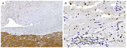
- Experimental details
- Figure from citation: PRKAR1A IHC in a CNC-associated cardiac myxoma. A, A photomicrograph of the specimen at low power exhibits the robust reactivity of the adjacent normal myocardium with antibodies directed against PRKAR1A. B, A high-power photomicrograph exhibits the lack of reactivity seen in the neoplastic (myxoma) cells, although the intratumoral histiocytes are still strongly reactive. Dilution: 1:8000
- Validation comment
- IHC
