Explore
 Validate
 Learn
LearnA700-039-T
antibody from Invitrogen Antibodies
Targeting: IRF1
MAR
 Western blot Immunocytochemistry
 Immunoprecipitation Immunohistochemistry Flow cytometry Other assay
Antibody data
- Antibody Data
- Antigen structure
- References [0]
- Comments [0]
- Validations
- Western blot [1]
- Immunocytochemistry [1]
- Immunohistochemistry [2]
- Flow cytometry [1]
- Other assay [1]
Submit
Validation data
Reference
Comment
Report error
- Product number
- A700-039-T - Provider product page

- Provider
- Invitrogen Antibodies
- Product name
- IRF1 Recombinant Rabbit Monoclonal Antibody (BLR039F)
- Antibody type
- Monoclonal
- Antigen
- Other
- Reactivity
- Human
- Host
- Rabbit
- Isotype
- IgG
- Antibody clone number
- BLR039F
- Vial size
- 10 µL
- Concentration
- 250 µg/mL
- Storage
- 4° C
No comments: Submit comment
Supportive validation
- Submitted by
- Invitrogen Antibodies (provider)
- Main image
- Experimental details
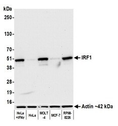
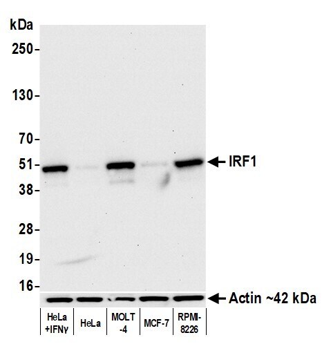
- Detection of human IRF1 by western blot. Samples: Whole cell lysate (10 µg) from HeLa treated with interferon gamma, HeLa, MOLT-4, MCF-7, and RPMI-8226 cells prepared using NETN lysis buffer. Antibody: Rabbit anti-IRF1 recombinant monoclonal antibody [BL-418-3E8] (Product # A700-039A lot 2) used at 1:1000. Secondary: HRP-conjugated goat anti-rabbit IgG (A120-101P). Chemiluminescence with an exposure time of 10 seconds. Lower Panel: Rabbit anti-Actin recombinant monoclonal antibody [BLR057F] (Product # A700-057).
Supportive validation
- Submitted by
- Invitrogen Antibodies (provider)
- Main image

- Experimental details
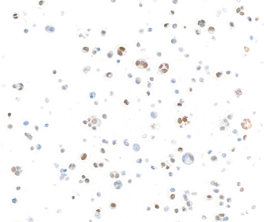
- Detection of human IRF1 by immunocytochemistry. Sample: FFPE section of MJ cells. Antibody: Rabbit anti-IRF1 recombinant monoclonal antibody (Product # A700-039 lot 2). Secondary: HRP-conjugated goat anti-rabbit IgG (A120-501P).
Supportive validation
- Submitted by
- Invitrogen Antibodies (provider)
- Main image
- Experimental details
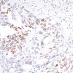
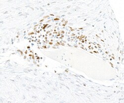
- Detection of human IRF1 by immunohistochemistry. AntibodySample: FFPE section of ovarian carcinoma.Antibody: Rabbit anti-IRF1 recombinant monoclonal antibody (BLR039F) (A700-039 lot 1) used at 1:250.Secondary: HRP-conjugated goat anti-rabbit IgG (A120-501P). Substrate: DAB.
- Submitted by
- Invitrogen Antibodies (provider)
- Main image
- Experimental details
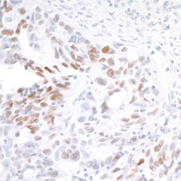
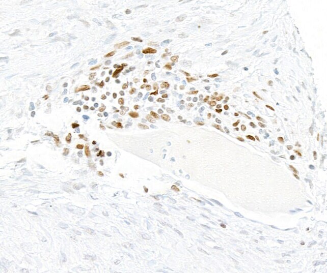
- Detection of human IRF1 by immunohistochemistry. Sample: FFPE section of ovarian carcinoma. Antibody: Rabbit anti-IRF1 recombinant monoclonal antibody (Product # A700-039 lot 2). Secondary: HRP-conjugated goat anti-rabbit IgG (A120-501P).
Supportive validation
- Submitted by
- Invitrogen Antibodies (provider)
- Main image
- Experimental details
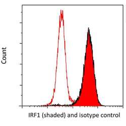
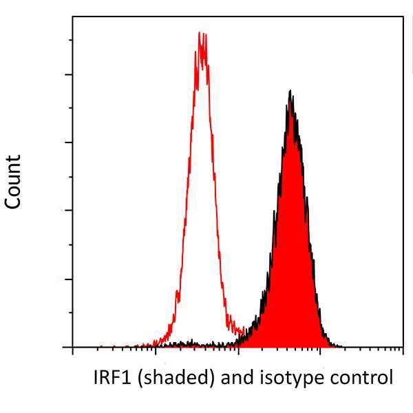
- Detection of human IRF1 (shaded) in MOLT4 cells by flow cytometry. Antibody: Rabbit anti-IRF1 recombinant monoclonal [BLR039F] (Product #A700-039; lot 1) or isotype control (unshaded). Secondary: DyLight® 650-conjugated goat anti-rabbit IgG (Product # A120-201D5).
Supportive validation
- Submitted by
- Invitrogen Antibodies (provider)
- Main image
- Experimental details
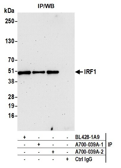
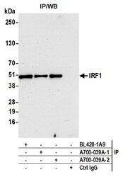
- Detection of human IRF1 by western blot of immunoprecipitates. Samples: Whole cell lysate (1.0 mg per IP reaction; 20% of IP loaded) from Jurkat cells prepared using NETN lysis buffer. Antibodies: Rabbit anti-IRF1 recombinant monoclonal antibody [BL-418-3E8] (Product # A700-039A lot 2) used for IP at 20 µL/mg lysate. IRF1 was also immunoprecipitated by a previous lot of this antibody (A700-039A lot 1) and a second antibody against a different epitope of IRF1 (BL428-1A9). For blotting immunoprecipitated IRF1 (Product # A700-039A) was used at 1:1000. Chemiluminescence with an exposure time of 75 seconds.