Explore
 Validate
 Learn
Learn Western blot
 ELISA
ELISAAntibody data
- Antibody Data
- Antigen structure
- References [0]
- Comments [0]
- Validations
- Western blot [1]
- Immunocytochemistry [2]
- Immunohistochemistry [10]
- Flow cytometry [1]
Submit
Validation data
Reference
Comment
Report error
- Product number
- RQ7634 - Provider product page

- Provider
- NSJ Bioreagents
- Product name
- SF3a60 Antibody / SAP 61 / Splicing factor 3A subunit 3
- Antibody type
- Polyclonal
- Description
- Antigen affinity purified
- Reactivity
- Human, Mouse, Rat
- Host
- Rabbit
- Vial size
- 100 µg
- Concentration
- 0.5mg/ml if reconstituted with 0.2ml sterile DI water
- Storage
- After reconstitution, the SF3a60 antibody can be stored for up to one month at 4oC. For long-term, aliquot and store at -20oC. Avoid repeated freezing and thawing.
No comments: Submit comment
Supportive validation
- Submitted by
- NSJ Bioreagents (provider)
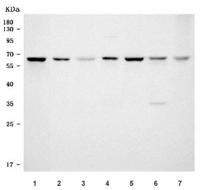
- Main image

- Experimental details
- Western blot testing of 1) human HeLa, 2) human A431, 3) human HL60, 4) rat liver, 5) rat C6, 6) mouse liver and 7) mouse NIH 3T3 cell lysate with SF3a60 antibody. Predicted molecular weight ~59 kDa.
Supportive validation
- Submitted by
- NSJ Bioreagents (provider)
- Main image
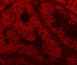
- Experimental details
- Immunofluorescent staining of FFPE human intestinal cancer tissue with SF3a60 antibody. HIER: steam section in pH8 EDTA buffer for 20 min.

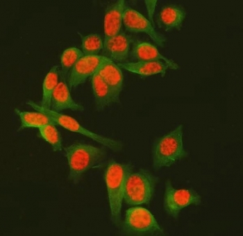
- Submitted by
- NSJ Bioreagents (provider)
- Main image
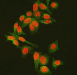
- Experimental details
- Immunofluorescent staining of FFPE human PC-3 cells with SF3a60 antibody (red) and Alpha Tubulin mAb (green). HIER: steam section in pH6 citrate buffer for 20 min.
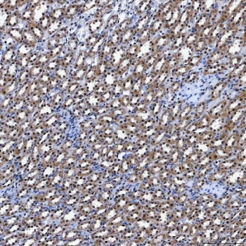
Supportive validation
- Submitted by
- NSJ Bioreagents (provider)
- Main image
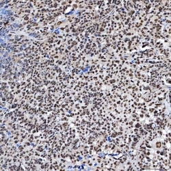
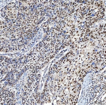
- Experimental details
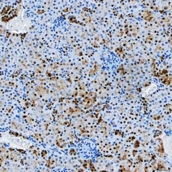
- IHC staining of FFPE human ovarian serous cancer tissue with SF3a60 antibody. HIER: boil tissue sections in pH8 EDTA for 20 min and allow to cool before testing.

- Submitted by
- NSJ Bioreagents (provider)
- Main image
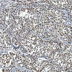
- Experimental details
- IHC staining of FFPE human breast cancer tissue with SF3a60 antibody. HIER: boil tissue sections in pH8 EDTA for 20 min and allow to cool before testing.

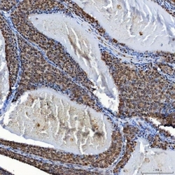
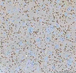
- Submitted by
- NSJ Bioreagents (provider)
- Main image
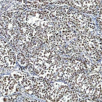
- Experimental details
- IHC staining of FFPE human larynx squamous cell carcinoma tissue with SF3a60 antibody. HIER: boil tissue sections in pH8 EDTA for 20 min and allow to cool before testing.
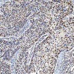
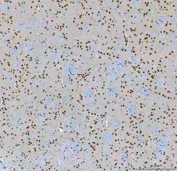
- Submitted by
- NSJ Bioreagents (provider)
- Main image
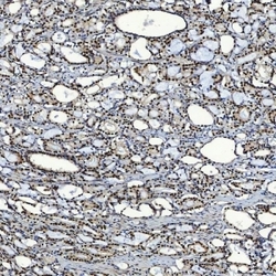
- Experimental details
- IHC staining of FFPE human lung adenocarcinoma tissue with SF3a60 antibody. HIER: boil tissue sections in pH8 EDTA for 20 min and allow to cool before testing.
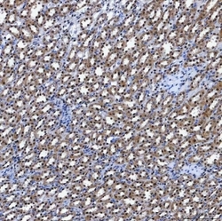
- Submitted by
- NSJ Bioreagents (provider)
- Main image
- Experimental details
- IHC staining of FFPE human prostate adenocarcinoma tissue with SF3a60 antibody. HIER: boil tissue sections in pH8 EDTA for 20 min and allow to cool before testing.
- Submitted by
- NSJ Bioreagents (provider)
- Main image

- Experimental details
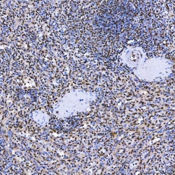
- IHC staining of FFPE human spleen tissue with SF3a60 antibody. HIER: boil tissue sections in pH8 EDTA for 20 min and allow to cool before testing.
- Submitted by
- NSJ Bioreagents (provider)
- Main image
- Experimental details
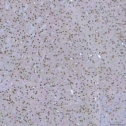
- IHC staining of FFPE mouse brain tissue with SF3a60 antibody. HIER: boil tissue sections in pH8 EDTA for 20 min and allow to cool before testing.
- Submitted by
- NSJ Bioreagents (provider)
- Main image
- Experimental details
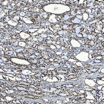
- IHC staining of FFPE mouse kidney tissue with SF3a60 antibody. HIER: boil tissue sections in pH8 EDTA for 20 min and allow to cool before testing.

- Submitted by
- NSJ Bioreagents (provider)
- Main image
- Experimental details
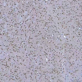
- IHC staining of FFPE rat brain tissue with SF3a60 antibody. HIER: boil tissue sections in pH8 EDTA for 20 min and allow to cool before testing.
- Submitted by
- NSJ Bioreagents (provider)
- Main image
- Experimental details
- IHC staining of FFPE rat kidney tissue with SF3a60 antibody. HIER: boil tissue sections in pH8 EDTA for 20 min and allow to cool before testing.
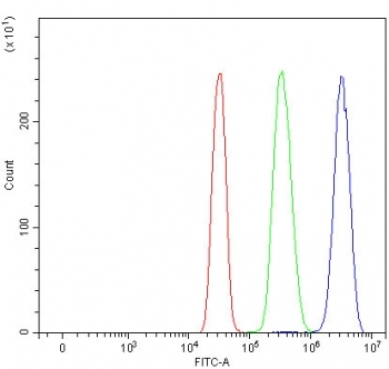
Supportive validation
- Submitted by
- NSJ Bioreagents (provider)
- Main image
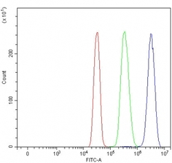
- Experimental details
- Flow cytometry testing of human SiHa cells with SF3a60 antibody at 1ug/million cells (blocked with goat sera); Red=cells alone, Green=isotype control, Blue= SF3a60 antibody.