Explore
 Validate
 Learn
Learn Western blot
Antibody data
- Antibody Data
- Antigen structure
- References [4]
- Comments [0]
- Validations
- Western blot [1]
- Immunohistochemistry [1]
- Other assay [2]
Submit
Validation data
Reference
Comment
Report error
- Product number
- PA1-86737 - Provider product page

- Provider
- Invitrogen Antibodies
- Product name
- SLUG Polyclonal Antibody
- Antibody type
- Polyclonal
- Antigen
- Synthetic peptide
- Description
- In Western blot, this antibody detects a band at ~30kDa in 293 cell lysate. Rabbit anti Slug antibody detects Slug, a member of the Snail family of zinc finger transcription factors, whose expression is induced by FGF, BMP, and TGF-beta.
- Reactivity
- Human, Mouse
- Host
- Rabbit
- Isotype
- IgG
- Vial size
- 100 µg
- Concentration
- 1 mg/mL
- Storage
- Store at 4°C short term. For long term storage, store at -20°C, avoiding freeze/thaw cycles.
Submitted references Chondroitin polymerizing factor promotes breast carcinoma cell proliferation, invasion and migration and affects expression of epithelial-mesenchymal transition-related markers.
PIK3CA Is Regulated by CUX1, Promotes Cell Growth and Metastasis in Bladder Cancer via Activating Epithelial-Mesenchymal Transition.
A Sox2-Sox9 signalling axis maintains human breast luminal progenitor and breast cancer stem cells.
Calreticulin Is Required for TGF-β-Induced Epithelial-to-Mesenchymal Transition during Cardiogenesis in Mouse Embryonic Stem Cells.
Li Y, Gong H, Feng L, Mao D, Xiao Y, Wang Y, Huang L
FEBS open bio 2021 Feb;11(2):423-434
FEBS open bio 2021 Feb;11(2):423-434
PIK3CA Is Regulated by CUX1, Promotes Cell Growth and Metastasis in Bladder Cancer via Activating Epithelial-Mesenchymal Transition.
Wang Z, Shang J, Li Z, Li H, Zhang C, He K, Li S, Ju W
Frontiers in oncology 2020;10:536072
Frontiers in oncology 2020;10:536072
A Sox2-Sox9 signalling axis maintains human breast luminal progenitor and breast cancer stem cells.
Domenici G, Aurrekoetxea-Rodríguez I, Simões BM, Rábano M, Lee SY, Millán JS, Comaills V, Oliemuller E, López-Ruiz JA, Zabalza I, Howard BA, Kypta RM, Vivanco MD
Oncogene 2019 Apr;38(17):3151-3169
Oncogene 2019 Apr;38(17):3151-3169
Calreticulin Is Required for TGF-β-Induced Epithelial-to-Mesenchymal Transition during Cardiogenesis in Mouse Embryonic Stem Cells.
Karimzadeh F, Opas M
Stem cell reports 2017 May 9;8(5):1299-1311
Stem cell reports 2017 May 9;8(5):1299-1311
No comments: Submit comment
Supportive validation
- Submitted by
- Invitrogen Antibodies (provider)
- Main image
- Experimental details
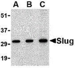
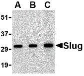
- Western blot analysis of whole cell lysate from HEK293 human epithelial kidney cells probed with a SLUG polyclonal antibody (Product # PA1-86737) at 0.5 (A), and 1 (B) and 2 (C) µg/mL
Supportive validation
- Submitted by
- Invitrogen Antibodies (provider)
- Main image

- Experimental details
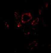
- Immunofluorescent analysis of human kidney using a SLUG polyclonal antibody (Product # PA1-86737).
Supportive validation
- Submitted by
- Invitrogen Antibodies (provider)
- Main image
- Experimental details
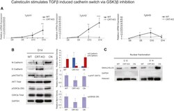
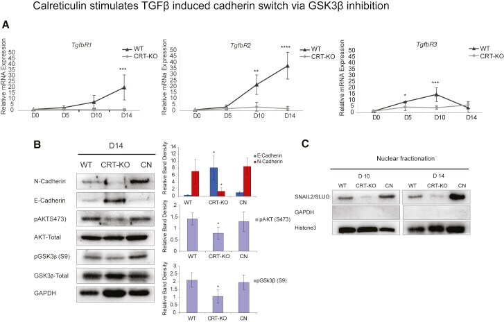
- Figure 3 Calreticulin Absence Reduces S9 Phosphorylation of GSK3beta and Affects SNAIL2/SLUG Nuclear Translocation (A) qPCR analysis shows the mRNA expression of TGF-beta receptor markers TgfbR1 , TgfbR2 , and TgfbR3 in WT and CRT-KO cells during differentiation. (B) N-cadherin, E-cadherin, pAKT (S473), and pGSK3beta (S9) versus total GSK3beta and total AKT were examined in WT, CRT-KO, and CN cells at D14 of cardiac differentiation from total cell lysates by western blot analysis. Compared with calreticulin-containing WT cells, in KO cells E-cadherin is more abundant, while in contrast N-cadherin is more abundant in WT cells than in calreticulin-deficient KO cells. CN cells express less E-cadherin and more N-cadherin. AKT phosphorylation at serine 473 and GSK3beta phosphorylation at S9 is low in CRT-KO cells. The bar graphs show the quantification of E-cad/N-cad, pAKT, and pGSK3beta band density versus GAPDH from three independent experiments; * p
- Submitted by
- Invitrogen Antibodies (provider)
- Main image
- Experimental details
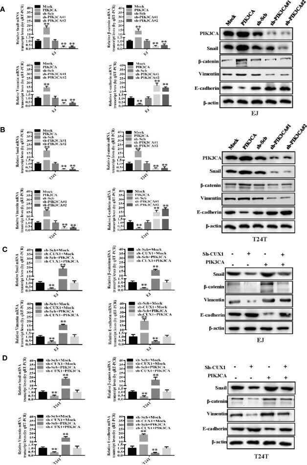
- Figure 5 PIK3CA promoted bladder cancer progression by activating EMT related makers--Snail, beta-catenin, vimentin and E-cadherin. (A, B) The mRNA expression and protein levels of Snail, beta-catenin, vimentin and E-cadherin were determined using RT-PCR and Western blot, respectively, in EJ or T24T cells stably transfected with mock, PIK3CA , sh-Scb and sh- PIK3CA . (C, D) The transcript and protein levels of Snail, beta-catenin, vimentin and E-cadherin in EJ or T24T cells stably transfected with mock, CUX1 , sh-Scb, and sh- CUX1 , and those co-transfected with PIK3CA and sh- PIK3CA , as detected by quantitative real-time PCR and Western blot. ** P < 0.01.