 Explore
 Validate
 Learn
Learn Western blot
 Immunoprecipitation
Antibody data
- Antibody Data
- Antigen structure
- References [4]
- Comments [0]
- Validations
- Western blot [2]
- Immunohistochemistry [1]
Submit
Validation data
Reference
Comment
Report error
- Product number
- NB100-2277 - Provider product page

- Provider
- Novus Biologicals
- Proper citation
- Novus Cat#NB100-2277, RRID:AB_2182065
- Product name
- Rabbit Polyclonal RPL7A Antibody
- Antibody type
- Polyclonal
- Description
- Immunogen affinity purified.
- Reactivity
- Human, Mouse
- Host
- Rabbit
- Isotype
- IgG
- Vial size
- 0.1 ml
- Concentration
- 0.2 mg/ml
- Storage
- Store at 4C. Do not freeze.
Submitted references Tracking the Fragile X Mental Retardation Protein in a Highly Ordered Neuronal RiboNucleoParticles Population: A Link between Stalled Polyribosomes and RNA Granules.
Molecular dynamics of FMRP and other RNA-binding proteins in MEG-01 differentiation: the role of mRNP complexes in non-neuronal development.
Unusual subcellular confinement of the fragile X mental retardation protein (FMRP) in circulating human platelets: complete polyribosome dissociation.
Functional Sindbis virus replicative complexes are formed at the plasma membrane.
El Fatimy R, Davidovic L, Tremblay S, Jaglin X, Dury A, Robert C, De Koninck P, Khandjian EW
PLoS genetics 2016 Jul;12(7):e1006192
PLoS genetics 2016 Jul;12(7):e1006192
Molecular dynamics of FMRP and other RNA-binding proteins in MEG-01 differentiation: the role of mRNP complexes in non-neuronal development.
McCoy M, Poliquin-Duchesneau D, Corbin F
Biochemistry and cell biology = Biochimie et biologie cellulaire 2016 Dec;94(6):597-608
Biochemistry and cell biology = Biochimie et biologie cellulaire 2016 Dec;94(6):597-608
Unusual subcellular confinement of the fragile X mental retardation protein (FMRP) in circulating human platelets: complete polyribosome dissociation.
Lauzière V, Lessard M, Meunier AJ, McCoy M, Bergeron LJ, Corbin F
Biochimie 2012 Apr;94(4):1069-73
Biochimie 2012 Apr;94(4):1069-73
Functional Sindbis virus replicative complexes are formed at the plasma membrane.
Frolova EI, Gorchakov R, Pereboeva L, Atasheva S, Frolov I
Journal of virology 2010 Nov;84(22):11679-95
Journal of virology 2010 Nov;84(22):11679-95
No comments: Submit comment
Supportive validation
- Submitted by
- Novus Biologicals (provider)
- Main image

- Experimental details
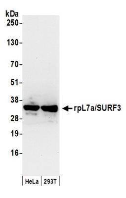
- Western Blot: RPL7A Antibody [NB100-2277] - Whole cell lysate (50 ug) from HeLa and 293T cells prepared using NETN lysis buffer. Antibody: Affinity purified rabbit anti-rpL7a/SURF3 antibody NB100-2277 used for WB at 0.1 ug/ml. Detection: Chemiluminescence with an exposure time of 10 seconds.
- Submitted by
- Novus Biologicals (provider)
- Main image

- Experimental details
- Western Blot: RPL7A Antibody [NB100-2277] - Detection of Human rpL7a (SURF3) on HeLa whole cell lysate using NB100-2277.

Supportive validation
- Submitted by
- Novus Biologicals (provider)
- Main image

- Experimental details
- Immunohistochemistry-Paraffin: RPL7A Antibody [NB100-2277] - Sample: FFPE sections of human colon carcinoma (left) and mouse renal cell carcinoma (right). Antibody: Affinity purified rabbit anti- rpL7a (SURF3) used at a dilution of 1:200 (1ug/ml). Detection: DAB
