 Explore
 Validate
 Learn
Learn Western blot
 Immunocytochemistry
 Immunoprecipitation
Antibody data
- Antibody Data
- Antigen structure
- References [0]
- Comments [0]
- Validations
- Immunocytochemistry [5]
- Immunohistochemistry [3]
- Flow cytometry [3]
Submit
Validation data
Reference
Comment
Report error
- Product number
- PA1-31405 - Provider product page

- Provider
- Invitrogen Antibodies
- Product name
- SR-BI Polyclonal Antibody
- Antibody type
- Polyclonal
- Antigen
- Synthetic peptide
- Description
- PA1-31405 detects SR-BI from rat, mouse, human samples.
- Reactivity
- Human, Mouse, Rat, Hamster, Rabbit
- Host
- Rabbit
- Isotype
- IgG
- Vial size
- 100 μL
- Concentration
- 1.0 mg/mL
- Storage
- 4°C, do not freeze
No comments: Submit comment
Supportive validation
- Submitted by
- Invitrogen Antibodies (provider)
- Main image

- Experimental details
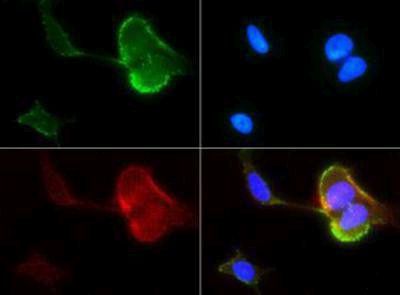
- Immunofluorescent analysis of SR-BI in HeLa cells using a SR-BI polyclonal antibody (Product # PA1-31405) at a dilution of 1:50 (green). Nuclei and actin were counterstained with DAPI (blue) and a Phalloidin (red).
- Submitted by
- Invitrogen Antibodies (provider)
- Main image

- Experimental details
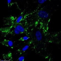
- Immunocytochemistry-Immunofluorescence analysis of SR-BI in human fibroblasts. Cells were fixed with 4% PFA, permeabilized with 0.2% Tween and SR-BI Polyclonal Antibody (Product # PA1-31405) (Green) at a dilution of 1:100 was used as a primary antibody. Blue: DAPI.

- Submitted by
- Invitrogen Antibodies (provider)
- Main image

- Experimental details
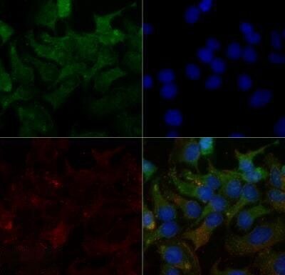
- Immunocytochemistry-Immunofluorescence analysis of SR-BI in HeLa cells using SR-BI Polyclonal Antibody (Product # PA1-31405) (Green) at a dilution of 2 µg/mL. Red : Tubulin. Blue: DAPI.
- Submitted by
- Invitrogen Antibodies (provider)
- Main image
- Experimental details
- Immunocytochemistry-Immunofluorescence analysis of SR-BI in human fibroblasts. Cells were fixed with 4% PFA, permeabilized with 0.2% Tween and SR-BI Polyclonal Antibody (Product # PA1-31405) (Green) at a dilution of 1:100 was used as a primary antibody. Blue: DAPI.

- Submitted by
- Invitrogen Antibodies (provider)
- Main image
- Experimental details
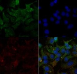
- Immunocytochemistry-Immunofluorescence analysis of SR-BI in HeLa cells using SR-BI Polyclonal Antibody (Product # PA1-31405) (Green) at a dilution of 2 µg/mL. Red : Tubulin. Blue: DAPI.

Supportive validation
- Submitted by
- Invitrogen Antibodies (provider)
- Main image
- Experimental details
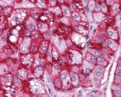
- Immunohistochemistry (Paraffin) analysis of SR-BI in human adrenal cortex tissue using SR-BI Polyclonal Antibody (Product # PA1-31405).

- Submitted by
- Invitrogen Antibodies (provider)
- Main image
- Experimental details
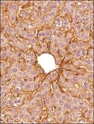
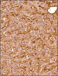
- Immunohistochemistry (Paraffin) analysis of SR-BI in mouse liver tissue using SR-BI Polyclonal Antibody (Product # PA1-31405) at a dilution of 1:300.

- Submitted by
- Invitrogen Antibodies (provider)
- Main image
- Experimental details
- Immunohistochemistry (Paraffin) analysis of SR-BI in mouse liver tissue using SR-BI Polyclonal Antibody (Product # PA1-31405) at a dilution of 1:300.

Supportive validation
- Submitted by
- Invitrogen Antibodies (provider)
- Main image
- Experimental details
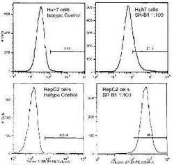
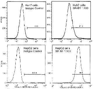
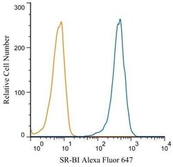
- Flow cytometry analysis of SR-BI in HepG2 cells using a SR-BI polyclonal antibody (Product # PA1-31405).
- Submitted by
- Invitrogen Antibodies (provider)
- Main image

- Experimental details
- Flow Cytometry analysis of TLR7 was performed in HeLa cells (fixed with 4% PFA, permeabilized with 0.1% saponin) using a TLR7 Polyclonal Antibody (Product # PA1-28109) (Blue) at a dilution of 2 µg/mL. Orange : isotype control.

- Submitted by
- Invitrogen Antibodies (provider)
- Main image
- Experimental details
- Flow Cytometry analysis of TLR7 was performed in HeLa cells (fixed with 4% PFA, permeabilized with 0.1% saponin) using a TLR7 Polyclonal Antibody (Product # PA1-28109) (Blue) at a dilution of 2 µg/mL. Orange : isotype control.
