Antibody data
- Antibody Data
- Antigen structure
- References [3]
- Comments [0]
- Validations
- Western blot [1]
- Immunocytochemistry [1]
- Immunohistochemistry [1]
- Other assay [3]
Submit
Validation data
Reference
Comment
Report error
- Product number
- MA1-204 - Provider product page

- Provider
- Invitrogen Antibodies
- Product name
- SFTPB Monoclonal Antibody (SPB02 (1B9))
- Antibody type
- Monoclonal
- Antigen
- Recombinant full-length protein
- Description
- Detects mature surfactant protein B (8kDa monomer and 16kDa Dimeric forms) and pro surfactant protein B (24kDa) in western blot. Sequence homology: Cow: 86%; Dog: 100%; Guinea Pig: 100%; Human: 100%; Mouse: 92%; Rabbit: 85%; Rat: 100%; Sheep: 79%.
- Reactivity
- Human
- Host
- Mouse
- Isotype
- IgG
- Antibody clone number
- SPB02 (1B9)
- Vial size
- 100 µg
- Concentration
- 1 mg/mL
- Storage
- -20° C, Avoid Freeze/Thaw Cycles
Submitted references Organoid-based expansion of patient-derived primary alveolar type 2 cells for establishment of alveolus epithelial Lung-Chip cultures.
Human Lung Stem Cell-Based Alveolospheres Provide Insights into SARS-CoV-2-Mediated Interferon Responses and Pneumocyte Dysfunction.
Abnormalities of the TITF-1 lineage-specific oncogene in NSCLC: implications in lung cancer pathogenesis and prognosis.
van Riet S, van Schadewijk A, Khedoe PPSJ, Limpens RWAL, Bárcena M, Stolk J, Hiemstra PS, van der Does AM
American journal of physiology. Lung cellular and molecular physiology 2022 Apr 1;322(4):L526-L538
American journal of physiology. Lung cellular and molecular physiology 2022 Apr 1;322(4):L526-L538
Human Lung Stem Cell-Based Alveolospheres Provide Insights into SARS-CoV-2-Mediated Interferon Responses and Pneumocyte Dysfunction.
Katsura H, Sontake V, Tata A, Kobayashi Y, Edwards CE, Heaton BE, Konkimalla A, Asakura T, Mikami Y, Fritch EJ, Lee PJ, Heaton NS, Boucher RC, Randell SH, Baric RS, Tata PR
Cell stem cell 2020 Dec 3;27(6):890-904.e8
Cell stem cell 2020 Dec 3;27(6):890-904.e8
Abnormalities of the TITF-1 lineage-specific oncogene in NSCLC: implications in lung cancer pathogenesis and prognosis.
Tang X, Kadara H, Behrens C, Liu DD, Xiao Y, Rice D, Gazdar AF, Fujimoto J, Moran C, Varella-Garcia M, Lee JJ, Hong WK, Wistuba II
Clinical cancer research : an official journal of the American Association for Cancer Research 2011 Apr 15;17(8):2434-43
Clinical cancer research : an official journal of the American Association for Cancer Research 2011 Apr 15;17(8):2434-43
No comments: Submit comment
Supportive validation
- Submitted by
- Invitrogen Antibodies (provider)
- Main image

- Experimental details
- Western blot analysis of Surfactant Protein B was performed by loading 20 µg of the indicated whole cell lysates and 5 µL of PageRuler Plus Prestained Protein Ladder (Product # 26619) per well onto a Novex 4-20% Tris-Glycine polyacrylamide gel (Product # WT4202BOX ). Proteins were transferred to a nitrocellulose membrane using the G2 Blotter (Product # 62288), and blocked with 5% Milk in TBST for 1 hour at room temperature. Mature Surfactant Protein B dimer was detected at ~16 kDa and Pro-Surfactant B at ~24 kDa using a Surfactant Protein B monoclonal antibody (Product # MA1-204) at a dilution of 2 µg/mL in 5% Milk in TBST overnight at 4C on a rocking platform, followed by a Goat anti-Mouse IgG (H+L) Superclonal™ Secondary Antibody, HRP conjugate (Product # A28177) at a dilution of 1:2,000 for at least 1 hour at room temperature. Chemiluminescent detection was performed using SuperSignal Pico substrate (Product # 34078) and the myECL Imager (Product # 62236).
Supportive validation
- Submitted by
- Invitrogen Antibodies (provider)
- Main image
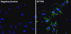
- Experimental details
- Immunofluorescent analysis of Surfactant Protein B (green) in A549 cells. The cells were fixed with 4% paraformaldehyde for 15 minutes, permeabilized with 0.1% Triton X-100 in PBS for 15 minutes, and blocked with 3% BSA in PBS (Product # 37525) for 30 minutes at room temperature. Cells were stained with a Surfactant Protein B monoclonal antibody (Product # MA1-204) at a dilution of 1:50 in staining buffer for 1 hour at room temperature, and then incubated with a Goat anti-Mouse IgG Superclonal™ Secondary Antibody, Alexa Fluor 488 conjugate (Product # A28175) at a dilution of 1:1000 for 1 hour at room temperature (green). Nuclei (blue) were counterstained with Hoechst 33342 dye (Product # 62249. Images were taken on a Thermo Scientific ToxInsight Instrument at 20X magnification.
Supportive validation
- Submitted by
- Invitrogen Antibodies (provider)
- Main image
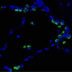
- Experimental details
- Immunofluorescent analysis of Mature Surfactant Protein B (green) in frozen human lung tissue. The cells were fixed with 4% paraformaldehyde for 5 minutes, and then antigen retrieval was performed using 10mM citrate buffer. Sections were blocked with 4% donkey serµm/PBST for 2hours at room temperature. Cells were stained with a Surfactant Protein B monoclonal antibody (Product # MA1-204) at a dilution of 1:100 in staining buffer overnight at 4C, and then incubated with a Goat anti-Mouse IgG Secondary Antibody at a dilution of 1:200 for 1 hour at room temperature (green). Nuclei (blue) were counterstained with DAPI. Images were taken on a Nikon A1Rsi inverted confocal microscope at 60x magnification.
Supportive validation
- Submitted by
- Invitrogen Antibodies (provider)
- Main image
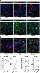
- Experimental details
- Figure 4. Alveolar type 2 cell culture after organoid-based expansion on insert and Alveolus Lung-Chip platform. Organoids from donors at different passages were dissociated, mixed and seeded on inserts or the Alveolus Lung-Chip and cultured for 7 days. A : fluorescent images of AEC2s after 7 days of culture on inserts or chips, in presence or absence of stretch. Cells were fixed and stained for HTII-280 (AEC2s, green), prosurfactant protein C (SP-C, red), and nuclei with DAPI (blue). Representative image from n = 4 independent experiments with different donor (mixes). B : the same cultures represented in A were furthermore stained with zonula occludens-1 (ZO-1, green), or C : surfactant proteins A (SP-A; red) or B (SP-B; green). D : at day 7 , culture cells were lysed and RNA was isolated followed by cDNA synthesis to assess gene expression levels of surfactant protein B ( SFTPB ) and C ( SFTPC ). Data are shown as delta Ct normalized for the geometric mean of the reference genes OAZ1 and ATP5B . Cultures on inserts are indicated with blue dots, chip control cultures with yellow dots and chip cultures exposed to 5 days of stretch with orange dots. The comparison between insert and chips was performed with three different donors. The comparison of stretch application with the control chip was performed with six different donor (mixes). AEC2s, alveolar type 2 cells.
- Submitted by
- Invitrogen Antibodies (provider)
- Main image
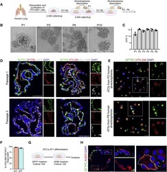
- Experimental details
- Figure 1 Establishment of Chemically Defined Human Lung Alveolosphere Culture System (A) Schematic representation of human alveolosphere cultures and passaging in SFFF medium. (B) Representative images of human alveolospheres from different passages. Scale bar: 100 mum. (C) Quantification of the colony formation efficiency (CFE) of human alveolospheres at different passages. (D) Immunostaining for SFTPC (green), SFTPB (red), and AGER (gray) (left panel) or SFTPB (green), HTII-280 (red), and DC-LAMP (gray) (right panel) at P1 and P3 human alveolospheres cultured in SFFF medium for 14 days. (E) Immunostaining for SFTPC (green) and HTII-280 (red) in cells dissociated from alveolospheres at P2 (top) and P8 (bottom). (F) Quantification of HTII-280 + SFTPC + cells/total 4',6-diamidino-2-phenylindole (DAPI) + cells derived from alveolospheres dissociation from P2 (orange) and P8 (blue). (G) Schematic representation of human AT2 to AT1 differentiation in alveolospheres. AT2s were cultured in SFFF medium for 10 days, followed by culture in ADM for 14 days. (H) Immunostaining for SFTPC (green) and AGER (red) in human alveolospheres cultured under ADM condition for 14 days. Scale bars: 100 mum (B); 50 mum (D); 20 mum (E); 20 mum (H). DAPI (blue) shows nuclei in (D), (E), and (H). Data are presented as mean +- SEM.
- Submitted by
- Invitrogen Antibodies (provider)
- Main image
- Experimental details
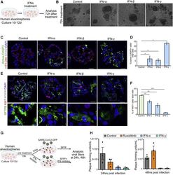
- Figure 6 IFN Treatment Recapitulates Features of SARS-CoV-2 Infection Including Cell Death and Loss of Surfactants in Alveolosphere-Derived AT2s (A) Schematic of experimental design. Human lung alveolospheres were treated with IFNalpha, IFNbeta or IFNgamma for 72 h. (B) Representative images of control and IFNalpha-, IFNbeta-, and IFNgamma-treated human lung alveolospheres. (C) Immunostaining for active-caspase-3 (green), HTII-280 (red), and SOX2 (gray) in control and IFN-treated alveolospheres. DAPI stains nuclei (blue). Scale bar: 30 mum. (D) Quantification of active caspase-3 + cells in total DAPI + (per alveolosphere) cells in control and IFN-treated human alveolospheres. (E) Immunostaining for SFTPB (green), Ki67 (red), and AGER (gray) in controls and IFNalpha-, IFNbeta-, or IFNgamma-treated human alveolospheres. DAPI stains nuclei (blue). Scale bar: 30 mum. (F) Quantification of Ki67 + cells in total DAPI + cells in control and IFN-treated human alveolospheres. * p < 0.05; ** p < 0.01; *** p < 0.001. (G) Schematic of IFNs or IFN inhibitor treatment followed by SARS-CoV-2 infection. (H) Viral titers in control (gray), ruxolitinib-treated (orange), IFNalpha-treated (blue), and IFNgamma-treated (green) cultures were determined by plaque assay using media collected from alveolosphere cultures at 24 and 48 h post-infection. Data are presented as mean +- SEM.
 Explore
 Validate
 Learn
Learn Western blot