Explore
 Validate
 Learn
Learn Western blot
 Immunoprecipitation
Antibody data
- Antibody Data
- Antigen structure
- References [0]
- Comments [0]
- Validations
- Western blot [1]
- Immunocytochemistry [1]
- Immunohistochemistry [2]
- Other assay [1]
Submit
Validation data
Reference
Comment
Report error
- Product number
- A700-069 - Provider product page

- Provider
- Invitrogen Antibodies
- Product name
- BRD3 Recombinant Rabbit Monoclonal Antibody (BLR069G)
- Antibody type
- Monoclonal
- Antigen
- Other
- Reactivity
- Human
- Host
- Rabbit
- Isotype
- IgG
- Antibody clone number
- BLR069G
- Vial size
- 100 µL
- Concentration
- 100 µg/mL
- Storage
- 4° C
No comments: Submit comment
Supportive validation
- Submitted by
- Invitrogen Antibodies (provider)
- Main image

- Experimental details
- Detection of human BRD3 by western blot. Samples: Whole cell lysate (50 µg) from HEK293T, RKO, Jurkat, LNCaP, HeLa, MCF-7, K-562, and Hep-G2 cells prepared using NETN lysis buffer. Antibody: Rabbit anti-BRD3 recombinant monoclonal antibody [BLR069G] (Product # A700-069 lot 1) used at 1:1000. Secondary: HRP-conjugated goat anti-rabbit IgG (A120-101P). Chemiluminescence with an exposure time of 10 seconds.

Supportive validation
- Submitted by
- Invitrogen Antibodies (provider)
- Main image

- Experimental details
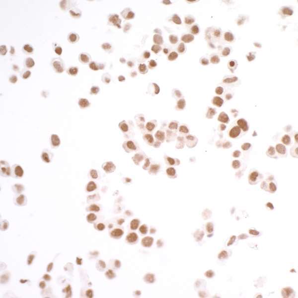
- Detection of human BRD3 by immunocytochemistry.Sample: FFPE section of OVCAR-3 cells.Antibody: Rabbit anti-BRD3 recombinant monoclonal antibody [BLR069G] (Product # A700-069; lot 1) used at 1:250.Secondary: HRP-conjugated goat anti-rabbit IgG (Product # A120-501P). Substrate: DAB.
Supportive validation
- Submitted by
- Invitrogen Antibodies (provider)
- Main image

- Experimental details
- Detection of human BRD3 by immunohistochemistry. AntibodySample: FFPE section of lung carcinoma.Antibody: Rabbit anti-BRD3 recombinant monoclonal antibody (BLR069G) (A700-069 lot 1) used at 1:250.Secondary: HRP-conjugated goat anti-rabbit IgG (A120-501P). Substrate: DAB.

- Submitted by
- Invitrogen Antibodies (provider)
- Main image
- Experimental details
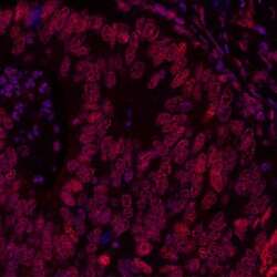
- Detection of human BRD3 by immunohistochemistry. Sample: FFPE section of human colon carcinoma. Antibody: Rabbit anti-BRD3 recombinant monoclonal antibody [BLR069G] (Product # A700-069 lot 1) used at 1:200. Secondary: DyLight® 594-conjugated goat anti-rabbit IgG (A120-101D4). Counterstain: DAPI (blue).

Supportive validation
- Submitted by
- Invitrogen Antibodies (provider)
- Main image
- Experimental details
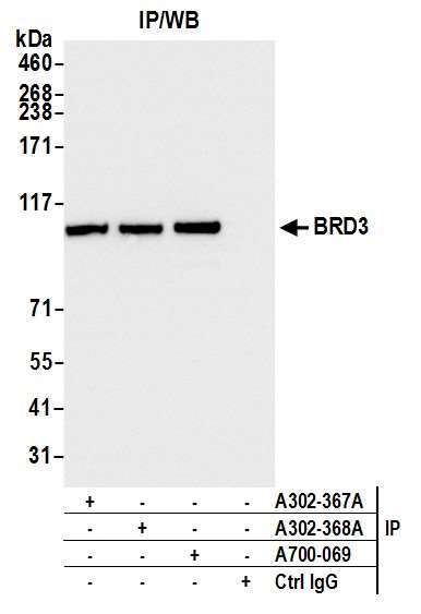
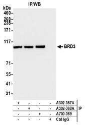
- Detection of human BRD3 by western blot of immunoprecipitates. Samples: Whole cell lysate (1.0 mg per IP reaction; 20% of IP loaded) from HEK293T cells prepared using NETN lysis buffer. Antibodies: Rabbit anti-BRD3 recombinant monoclonal antibody [BLR069G] (Product # A700-069 lot 1) used for IP at 20 µL/mg lysate. BRD3 was also immunoprecipitated by rabbit anti-BRD3 antibodies (Product# A302-367A and Product # A302-368A). For blotting immunoprecipitated BRD3 (Product # A700-069) was used at 1:1000. Chemiluminescence with an exposure time of 1 second.