 Explore
 Validate
 Learn
Learn Western blot
Antibody data
- Antibody Data
- Antigen structure
- References [0]
- Comments [0]
- Validations
- Western blot [2]
- Immunocytochemistry [1]
- Chromatin Immunoprecipitation [1]
Submit
Validation data
Reference
Comment
Report error
- Product number
- 703745 - Provider product page

- Provider
- Invitrogen Antibodies
- Product name
- WDR5 Recombinant Rabbit Monoclonal Antibody (9H25L13)
- Antibody type
- Monoclonal
- Antigen
- Other
- Description
- This antibody is predicted to react with Mouse, Bovine
- Reactivity
- Human, Rat
- Host
- Rabbit
- Isotype
- IgG
- Antibody clone number
- 9H25L13
- Vial size
- 100 µg
- Concentration
- 0.5 mg/mL
- Storage
- Store at 4°C short term. For long term storage, store at -20°C, avoiding freeze/thaw cycles.
No comments: Submit comment
Supportive validation
- Submitted by
- Invitrogen Antibodies (provider)
- Main image

- Experimental details
- Knockdown of WDR5 was achieved by transfecting HCT116 cells with WDR5 specific siRNA (Silencer® select Product # s21862 & s21863). Western blot analysis (Fig a) was performed using modified whole cell extracts (1% SDS) from WDR5 knockdown cells (Lane 3), non-specific scrambled siRNA transfected cells (Lane 2) and untransfected cells (Lane 1). The blot was probed with Anti-WDR5 Recombinant Rabbit Monoclonal Antibody (Product # 703475, 1:5000 dilution) and Goat anti-Rabbit IgG (H+L) Superclonal™ Secondary Antibody, HRP conjugate (Product # A27036, 1:5000 dilution). Densitometric analysis of this western blot is shown in the histogram (Fig b). Loss of signal upon siRNA mediated knockdown confirms that antibody is specific to WDR5.

- Submitted by
- Invitrogen Antibodies (provider)
- Main image

- Experimental details
- Western blot was performed using Anti-WDR5 Recombinant Rabbit Monoclonal Antibody (Product # 703475) and a 37 kDa band corresponding to WDR5 was observed across the cell lines tested. Modified whole cell extracts (1% SDS) (30 µg lysate) of HCT 116 (Lane 1), HT-29 (Lane 2), SW480 (Lane 3), COLO 205 (Lane 4), THP-1 (Lane 5) and PC-12 (Lane 6) were electrophoresed using Novex® NuPAGE® 10 % Bis-Tris gel (Product # NP0302BOX). Resolved proteins were then transferred onto a nitrocellulose membrane (Product # IB23001) by iBlot® 2 Dry Blotting System (Product # IB21001). The blot was probed with the primary antibody (1:5000 dilution) and detected by chemiluminescence Goat Anti-Rabbit IgG (H+L) Superclonal™ Secondary Antibody, HRP conjugate (Product # A27036, 1:5000 dilution). Chemiluminescent detection was performed using Novex® ECL Chemiluminescent Substrate Reagent Kit (Product # WP20005).

Supportive validation
- Submitted by
- Invitrogen Antibodies (provider)
- Main image

- Experimental details
- For immunofluorescence analysis, HCT 116 cells were fixed and permeabilized for detection of endogenous WDR5 using Anti-WDR5 Recombinant Rabbit Monoclonal Antibody (Product # 703475, 1:100 dilution) and labeled with Goat anti-Rabbit IgG (H+L) Highly Cross-Adsorbed Secondary Antibody, Alexa Fluor Plus 488 (Product # A32731, 1:2000). Panel a) shows representative cells that were stained for detection and localization of WDR5 protein (green), Panel b) is stained for nuclei (blue) using ProLong™ Diamond Antifade Mountant with DAPI (Product # P36962). Panel c) represents cytoskeletal F-actin staining using Rhodamine Phalloidin (Product # R415, 1:300). Panel d) is a composite image of Panels a, b and c clearly demonstrating nuclear localization of WDR5. Panel e) represents control cells with no primary antibody to assess background. The images were captured at 60X magnification.

Supportive validation
- Submitted by
- Invitrogen Antibodies (provider)
- Main image
- Experimental details
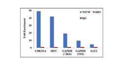
- Chromatin Immunoprecipitation (ChIP) assay of endogenous WDR5 protein using Anti-WDR5 Antibody: ChIP was performed using Anti-WDR5 Recombinant Rabbit monoclonal Antibody (Product # 703745, 5 µg) on sheared chromatin from 2 million HCT116 cells using the MAGnify ChIP System kit (Product # 49-2024). Normal Rabbit IgG was used as a negative IP control. The purified DNA was analyzed by qPCR using primers binding to CDKN1A intron1, c-MYC promoter, Gene body (+2Kb) and transcriptional start site of GAPDH and SAT2 satellite repeats. Data is presented as fold enrichment of the antibody signal versus the negative control IgG using the comparative CT method.
