Explore
 Validate
 Learn
LearnMA5-36184
antibody from Invitrogen Antibodies
Targeting: CHMP2B
CHMP2.5, DKFZP564O123, VPS2B
 Western blot Immunocytochemistry
 Immunoprecipitation Immunohistochemistry Flow cytometry Other assay
Antibody data
- Antibody Data
- Antigen structure
- References [0]
- Comments [0]
- Validations
- Western blot [3]
- Immunocytochemistry [5]
- Immunohistochemistry [2]
- Flow cytometry [1]
- Other assay [1]
Submit
Validation data
Reference
Comment
Report error
- Product number
- MA5-36184 - Provider product page

- Provider
- Invitrogen Antibodies
- Product name
- CHMP2B Recombinant Rabbit Monoclonal Antibody (JE54-35)
- Antibody type
- Monoclonal
- Antigen
- Recombinant full-length protein
- Reactivity
- Human, Mouse, Rat
- Host
- Rabbit
- Isotype
- IgG
- Antibody clone number
- JE54-35
- Vial size
- 100 µL
- Concentration
- 1 mg/mL
- Storage
- Store at 4°C short term. For long term storage, store at -20°C, avoiding freeze/thaw cycles.
No comments: Submit comment
Supportive validation
- Submitted by
- Invitrogen Antibodies (provider)
- Main image
- Experimental details
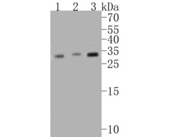
- Western blot analysis of CHMP2B using a CHMP2B monoclonal antibody (Product #MA5-36184). Proteins were transferred to a PVDF membrane and blocked with 5% BSA in PBS for 1 hour at room temperature. The primary antibody at a dilution of 1:500 was used in 5% BSA at room temperature for 2 hours. Goat Anti-Rabbit IgG - HRP Secondary Antibody at 1:5,000 dilution was used for 1 hour at room temperature. Positive control: Lane 1: Mouse bone marrow tissue lysate, Lane 2: Rat bone marrow tissue lysate, Lane 3: Human skeletal muscle tissue lysate

- Submitted by
- Invitrogen Antibodies (provider)
- Main image
- Experimental details
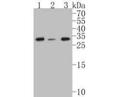
- Western blot analysis of CHMP2B using a CHMP2B monoclonal antibody (Product #MA5-36184). Proteins were transferred to a PVDF membrane and blocked with 5% BSA in PBS for 1 hour at room temperature. The primary antibody at a dilution of 1:500 was used in 5% BSA at room temperature for 2 hours. Goat Anti-Rabbit IgG - HRP Secondary Antibody at 1:5,000 dilution was used for 1 hour at room temperature. Positive control: Lane 1: A549 cell lysate, Lane 2: A431 cell lysate, Lane 3: Hela cell lysate

- Submitted by
- Invitrogen Antibodies (provider)
- Main image
- Experimental details
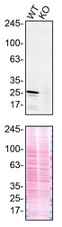
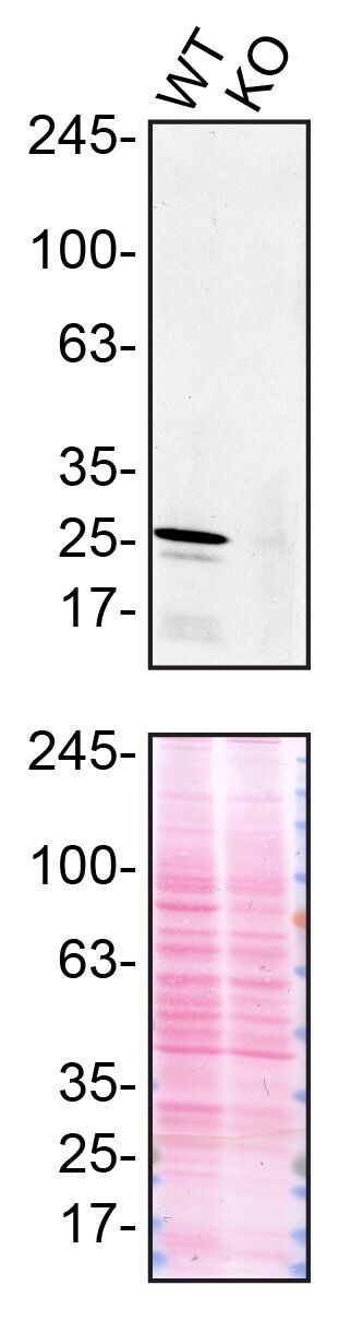
- Western blot of CHMP2B was performed by loading 50 µg of WT (lane 1) and CHMP2B CRISPR KO (lane 2) U2OS cell lysates in RIPA buffer onto a 4-20% gradient polyacrylamide gel. Proteins on the blots were visualized with Ponceau staining (below immunoblot). Proteins were transferred to nitrocellulose membrane and blocked in 5% milk for 1 hr. CHMP2B was detected at approximately 24 kDa using a CHMP2B recombinant monoclonal antibody (Product # MA5-36184) at a dilution of 1:500 in 5% BSA in TBS with 0.1% Tween 20 (TBST) overnight at 4°C. The peroxidase-conjugated secondary antibody (Product # 65-6120) was diluted to 0.2 µg/mL in TBST with 5% milk for 1 hr. Chemiluminescent detection was performed using Pierce ECL Western Blotting Substrate (Product # 32106). Data courtesy of YCharOS Inc., an open science company with the mission of characterizing commercially available antibodies using knockout validation.
Supportive validation
- Submitted by
- Invitrogen Antibodies (provider)
- Main image
- Experimental details
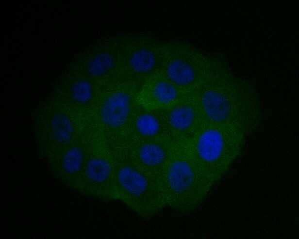
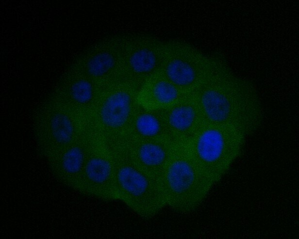
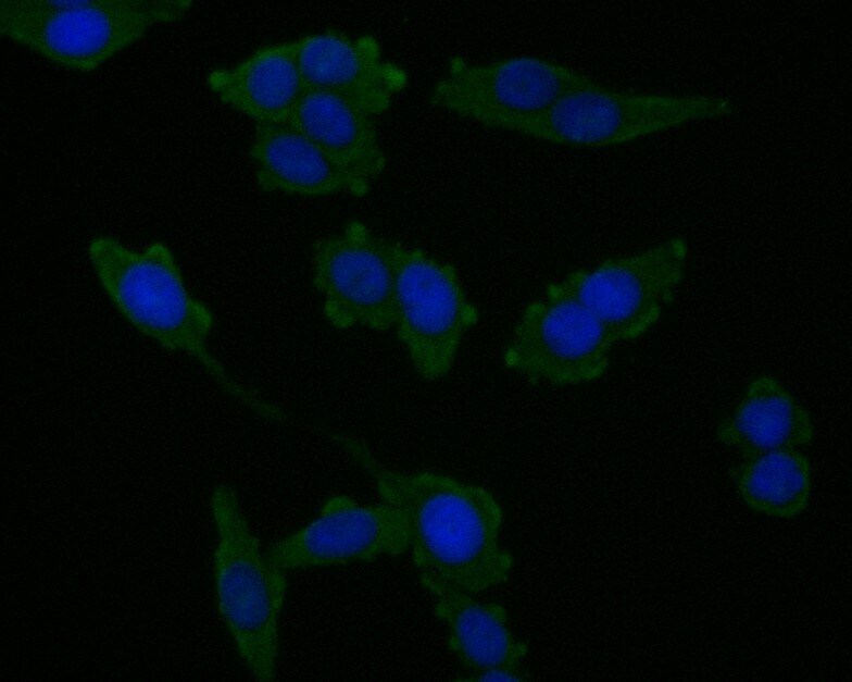
- Immunocytochemical analysis of CHMP2B in SW1990 cells using a CHMP2B monoclonal antibody (Product #MA5-36184). Formalin fixed cells were permeabilized with 0.1% Triton X-100 in TBS for 10 minutes at room temperature and blocked with 1% Blocker BSA for 15 minutes at room temperature. Cells were probed with the primary antibody for 1 hour at room temperature, washed with PBS. Alexa Fluor®488 Goat anti-Rabbit IgG was used as the secondary antibody at 1:1,000 dilution. The nuclear counter stain is DAPI.
- Submitted by
- Invitrogen Antibodies (provider)
- Main image

- Experimental details
- Immunocytochemical analysis of CHMP2B in SW620 cells using a CHMP2B monoclonal antibody (Product #MA5-36184). Formalin fixed cells were permeabilized with 0.1% Triton X-100 in TBS for 10 minutes at room temperature and blocked with 1% Blocker BSA for 15 minutes at room temperature. Cells were probed with the primary antibody for 1 hour at room temperature, washed with PBS. Alexa Fluor®488 Goat anti-Rabbit IgG was used as the secondary antibody at 1:1,000 dilution. The nuclear counter stain is DAPI.

- Submitted by
- Invitrogen Antibodies (provider)
- Main image

- Experimental details
- Immunocytochemical analysis of CHMP2B in SW1990 cells using a CHMP2B monoclonal antibody (Product #MA5-36184). Formalin fixed cells were permeabilized with 0.1% Triton X-100 in TBS for 10 minutes at room temperature and blocked with 1% Blocker BSA for 15 minutes at room temperature. Cells were probed with the primary antibody for 1 hour at room temperature, washed with PBS. Alexa Fluor®488 Goat anti-Rabbit IgG was used as the secondary antibody at 1:1,000 dilution. The nuclear counter stain is DAPI.
- Submitted by
- Invitrogen Antibodies (provider)
- Main image

- Experimental details
- Immunocytochemical analysis of CHMP2B in SW620 cells using a CHMP2B monoclonal antibody (Product #MA5-36184). Formalin fixed cells were permeabilized with 0.1% Triton X-100 in TBS for 10 minutes at room temperature and blocked with 1% Blocker BSA for 15 minutes at room temperature. Cells were probed with the primary antibody for 1 hour at room temperature, washed with PBS. Alexa Fluor®488 Goat anti-Rabbit IgG was used as the secondary antibody at 1:1,000 dilution. The nuclear counter stain is DAPI.
- Submitted by
- Invitrogen Antibodies (provider)
- Main image
- Experimental details
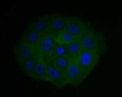
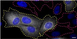
- Immunofluorescence of CHMP2B was performed using U2OS wild-type and CHMP2B KO cells that were transfected with a green or a far-red fluorescent dye, respectively. Post-transfection, WT and KO cells were mixed and plated to a 1:1 ratio on coverslips as a mosaic and incubated for 24 hrs. Cells were fixed in 4% PFA (in PBS) or methanol for 15 min; cells were permeabilized with 0.1% Triton X-100 for 10 min at RT and blocked with PBS with 5% BSA, 5% goat serum, and 0.01% Triton X-100 for 30 min. Cells were stained with the CHMP2B recombinant monoclonal antibody (Product # MA5-36184) at a 1:1,000 dilution overnight at 4°C. Secondary antibody incubation was performed using 1 µg/mL of Goat anti-Rabbit IgG (H+L) Highly Cross-Adsorbed Secondary Antibody, Alexa Fluor 555 antibody (Product # A21429) together with DAPI for 1 hr. Imaging was performed with a 40X oil objective and analysis was performed using Image J. Cell image represents a single focal plane; WT and KO cells are outlined with a yellow (WT) or magenta (KO) dashed line. Data courtesy of YCharOS Inc., an open science company with the mission of characterizing commercially available antibodies using knockout validation.

Supportive validation
- Submitted by
- Invitrogen Antibodies (provider)
- Main image
- Experimental details
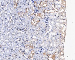
- Immunohistochemical analysis of CHMP2B in paraffin-embedded mouse kidney tissue using a monoclonal antibody (Product #MA5-36184). The section was pre-treated using heat mediated antigen retrieval with Tris-EDTA buffer (pH 8.0-8.4) for 20 minutes. The tissues were blocked in 5% BSA for 30 minutes at room temperature, washed with ddH2O and PBS, and then probed with the primary antibody (1:100) for 30 minutes at room temperature. The detection was performed using an HRP conjugated compact polymer system. DAB was used as the chromogen. Tissues were counterstained with hematoxylin and mounted with DPX.

- Submitted by
- Invitrogen Antibodies (provider)
- Main image

- Experimental details
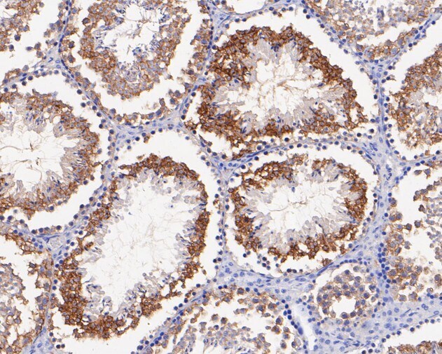
- Immunohistochemical analysis of CHMP2B in paraffin-embedded mouse testis tissue using a monoclonal antibody (Product #MA5-36184). The section was pre-treated using heat mediated antigen retrieval with Tris-EDTA buffer (pH 8.0-8.4) for 20 minutes. The tissues were blocked in 5% BSA for 30 minutes at room temperature, washed with ddH2O and PBS, and then probed with the primary antibody (1:100) for 30 minutes at room temperature. The detection was performed using an HRP conjugated compact polymer system. DAB was used as the chromogen. Tissues were counterstained with hematoxylin and mounted with DPX.
Supportive validation
- Submitted by
- Invitrogen Antibodies (provider)
- Main image
- Experimental details
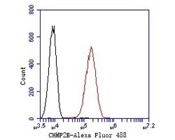
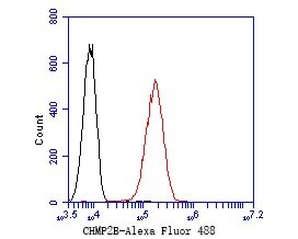
- Flow cytometric analysis of CHMP2B using A549 cells and a CHMP2B monoclonal antibody (Product #MA5-36184). The cells were fixed, permeabilized and stained with the primary antibody at a dilution of 1:50 (red). After incubation of the primary antibody at room temperature for an hour, the cells were stained with a Alexa Fluor 488-conjugated Goat anti-Rabbit IgG Secondary antibody at 1:1000 dilution for 30 minutes. Unlabeled sample was used as a control (cells without incubation with primary antibody; black).
Supportive validation
- Submitted by
- Invitrogen Antibodies (provider)
- Main image

- Experimental details
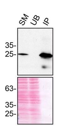
- Immunoprecipitation of CHMP2B was performed on U2OS cell lysates. Antibody-bead conjugates were prepared by adding 1 µg of CHMP2B recombinant monoclonal antibody (Product # MA5-36184) with 30 µL of protein A-Sepharose beads and rocked overnight at 4°C. 1 mg of lysate was incubated with an antibody-bead conjugate for 2 hours at 4°C. Following centrifugation and multiple washes, 10% starting material (SM), 10% unbound fraction (UB) and immunoprecipitated fraction (IP) were processed for immunoblot using a different CHMP2B recombinant monoclonal antibody. Ponceau stained transfer of blot is shown. Data courtesy of YCharOS Inc., an open science company with the mission of characterizing commercially available antibodies using knockout validation.