Explore
 Validate
 Learn
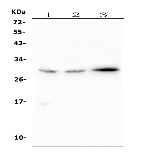
Learn Western blot
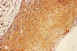
 Immunohistochemistry
Antibody data
- Antibody Data
- Antigen structure
- References [3]
- Comments [0]
- Validations
- Immunohistochemistry [1]
Submit
Validation data
Reference
Comment
Report error
- Product number
- PA1707 - Provider product page

- Provider
- Boster Biological Technology
- Product name
- Anti-SOCS3 Antibody
- Antibody type
- Polyclonal
- Description
- Polyclonal antibody for SOCS-3/SOCS3 detection. Host: Rabbit.Size: 100μg/vial. Tested applications: IHC-P. Reactive species: Human. SOCS-3/SOCS3 information: Molecular Weight: 24770 MW; Tissue Specificity: Widely expressed with high expression in heart, placenta, skeletal muscle, peripheral blood leukocytes, fetal and adult lung, and fetal liver and kidney. Lower levels in thymus.
- Reactivity
- Human, Mouse
- Host
- Rabbit
- Vial size
- 100μg/vial
- Concentration
- Add 0.2ml of distilled water will yield a concentration of 500ug/ml.
- Storage
- At -20°C for one year. After reconstitution, at 4°C for one month. It can also be aliquoted and stored frozen at -20°C for a longer time. Avoid repeated freezing and thawing.
- Handling
- Add 0.2ml of distilled water will yield a concentration of 500ug/ml.
Submitted references Glutathione-S-transferase A3 protein suppresses thiram-induced tibial dyschondroplasia by regulating prostaglandin-related genes expression.
The expression of prostaglandins-related genes in erythrocytes of broiler chicken responds to thiram-induced tibial dyschondroplasia and recombinant glutathione-S-transferase A3 protein.
Role of the JAK2/STAT3 signaling pathway in the pathogenesis of type 2 diabetes mellitus with macrovascular complications.
Niu S, Li X, Jahejo AR, Zhang N, Yang SX, Jia YF, Zhang YY, Tian ZX, Li Z, Ning GB, Zhang D, Tian WX
Research in veterinary science 2021 Mar;135:343-348
Research in veterinary science 2021 Mar;135:343-348
The expression of prostaglandins-related genes in erythrocytes of broiler chicken responds to thiram-induced tibial dyschondroplasia and recombinant glutathione-S-transferase A3 protein.
Niu S, Wang CX, Jia FJ, Jahejo AR, Li X, Ning GB, Zhang D, Ma HL, Hao WF, Gao WW, Zhao YJ, Gao SM, Li JH, Li GL, Yan F, Gao RK, Huo NR, Tian WX, Chen HC
Research in veterinary science 2019 Jun;124:112-117
Research in veterinary science 2019 Jun;124:112-117
Role of the JAK2/STAT3 signaling pathway in the pathogenesis of type 2 diabetes mellitus with macrovascular complications.
Yang M, Tian M, Zhang X, Xu J, Yang B, Yu J, Li F, Li Y, Li S, Li X
Oncotarget 2017 Nov 14;8(57):96958-96969
Oncotarget 2017 Nov 14;8(57):96958-96969
No comments: Submit comment
Supportive validation
- Submitted by
- Boster Biological Technology (provider)
- Main image
- Experimental details
- IHC analysis of SOCS3 using anti-SOCS3 antibody (PA1707). SOCS3 was detected in paraffin-embedded section of human tonsil tissues. Heat mediated antigen retrieval was performed in citrate buffer (pH6, epitope retrieval solution) for 20 mins. The tissue section was blocked with 10% goat serum. The tissue section was then incubated with 1μg/ml rabbit anti-SOCS3 Antibody (PA1707) overnight at 4°C. Biotinylated goat anti-rabbit IgG was used as secondary antibody and incubated for 30 minutes at 37°C. The tissue section was developed using Strepavidin-Biotin-Complex (SABC)(Catalog # SA1022) with DAB as the chromogen.
- Additional image