 Explore
 Validate
 Learn
LearnHPA042935
antibody from Atlas Antibodies
Targeting: CHCHD3
FLJ20420, Mic19, MICOS19, MINOS3, PPP1R22
 Western blot
Antibody data
- Antibody Data
- Antigen structure
- References [0]
- Comments [0]
- Validations
- Western blot [3]
- Immunocytochemistry [1]
- Immunohistochemistry [5]
Submit
Validation data
Reference
Comment
Report error
- Product number
- HPA042935 - Provider product page

- Provider
- Atlas Antibodies
- Proper citation
- Atlas Antibodies Cat#HPA042935, RRID:AB_10794538
- Product name
- Anti-CHCHD3
- Antibody type
- Polyclonal
- Reactivity
- Human, Mouse, Rat
- Host
- Rabbit
- Conjugate
- Unconjugated
- Antigen sequence
DENENITVVKGIRLSENVIDRMKESSPSGSKSQRY
SGAYGASVSDEELKRRVAEELALEQAKKESEDQKR
LKQAK- Isotype
- IgG
- Vial size
- 100 µl
- Storage
- Store at +4°C for short term storage. Long time storage is recommended at -20°C.
No comments: Submit comment
Supportive validation
- Submitted by
- Atlas Antibodies (provider)
- Enhanced method
- Genetic validation
- Main image

- Experimental details
- Western blot analysis in U-87MG ATCC cells transfected with control siRNA, target specific siRNA probe #1 and #2, using Anti-CHCHD3 antibody. Remaining relative intensity is presented. Loading control: Anti-GAPDH.

- Submitted by
- Atlas Antibodies (provider)
- Main image

- Experimental details
- Western blot analysis in human cell line HEK 293.

- Submitted by
- Atlas Antibodies (provider)
- Main image

- Experimental details
- Western blot analysis in mouse cell line NIH-3T3 and rat cell line NBT-II.

Supportive validation
- Submitted by
- Atlas Antibodies (provider)
- Main image

- Experimental details
- Immunofluorescent staining of human cell line U-2 OS shows localization to mitochondria.
- Sample type
- HUMAN

Supportive validation
- Submitted by
- Atlas Antibodies (provider)
- Main image

- Experimental details
- Immunohistochemical staining of human stomach, lower shows strong granular cytoplasmic positivity in glandular cells.

- Submitted by
- Atlas Antibodies (provider)
- Main image

- Experimental details
- Immunohistochemical staining of human kidney shows strong granular cytoplasmic positivity in cells in tubules.
- Sample type
- HUMAN

- Submitted by
- Atlas Antibodies (provider)
- Main image

- Experimental details
- Immunohistochemical staining of human duodenum shows strong granular cytoplasmic positivity in glandular cells.
- Sample type
- HUMAN

- Submitted by
- Atlas Antibodies (provider)
- Main image
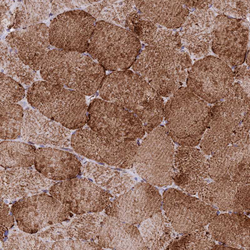
- Experimental details
- Immunohistochemical staining of human skeletal muscle shows strong granular cytoplasmic positivity in myocytes.
- Sample type
- HUMAN

- Submitted by
- Atlas Antibodies (provider)
- Main image

- Experimental details
- Immunohistochemical staining of human liver shows moderate granular cytoplasmic positivity in hepatocytes.
- Sample type
- HUMAN
