Explore
 Validate
 Learn
Learn Western blot
Antibody data
- Antibody Data
- Antigen structure
- References [1]
- Comments [0]
- Validations
- Western blot [1]
- Immunocytochemistry [1]
- Immunohistochemistry [4]
- Flow cytometry [1]
Submit
Validation data
Reference
Comment
Report error
- Product number
- TA501109 - Provider product page

- Provider
- OriGene
- Proper citation
- OriGene Cat#TA501109, RRID:AB_11124826
- Product name
- Anti-NIT2 mouse monoclonal antibody, clone OTI2B9 (formerly 2B9)
- Antibody type
- Monoclonal
- Description
- Anti-NIT2 mouse monoclonal antibody, clone OTI2B9 (formerly 2B9)
- Host
- Mouse
- Conjugate
- Unconjugated
- Epitope
- NIT2
- Isotype
- IgG
- Antibody clone number
- OTI2B9
- Vial size
- 100 µl
- Concentration
- 0.74 mg/ml
Submitted references ω-Amidase: an underappreciated, but important enzyme in L-glutamine and L-asparagine metabolism; relevance to sulfur and nitrogen metabolism, tumor biology and hyperammonemic diseases.
Cooper AJ, Shurubor YI, Dorai T, Pinto JT, Isakova EP, Deryabina YI, Denton TT, Krasnikov BF
Amino acids 2016 Jan;48(1):1-20
Amino acids 2016 Jan;48(1):1-20
No comments: Submit comment
Supportive validation
- Submitted by
- OriGene (provider)
- Main image
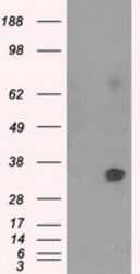
- Experimental details
- HEK293T cells were transfected with the pCMV6-ENTRY control (Left lane) or pCMV6-ENTRY NIT2 (RC210660, Right lane) cDNA for 48 hrs and lysed. Equivalent amounts of cell lysates (5 ug per lane) were separated by SDS-PAGE and immunoblotted with anti-NIT2.
- Validation comment
- WB
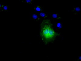
Supportive validation
- Submitted by
- OriGene (provider)
- Main image

- Experimental details
- Anti-NIT2 mouse monoclonal antibody (TA501109) immunofluorescent staining of COS7 cells transiently transfected by pCMV6-ENTRY NIT2(RC210660).
- Validation comment
- IF
Supportive validation
- Submitted by
- OriGene (provider)
- Main image
- Experimental details
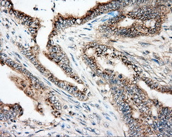
- Immunohistochemical staining of paraffin-embedded Adenocarcinoma of colon tissue using anti-NIT2 mouse monoclonal antibody. (Heat-induced epitope retrieval by 10mM citric buffer, pH6.0, 100C for 10min, TA501109, Dilution 1:50)
- Validation comment
- IHC

- Submitted by
- OriGene (provider)
- Main image
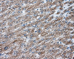
- Experimental details
- Immunohistochemical staining of paraffin-embedded liver tissue within the normal limits using anti-NIT2 mouse monoclonal antibody. (Heat-induced epitope retrieval by 10mM citric buffer, pH6.0, 100C for 10min, TA501109, Dilution 1:50)
- Validation comment
- IHC
- Submitted by
- OriGene (provider)
- Main image
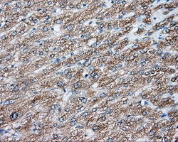
- Experimental details
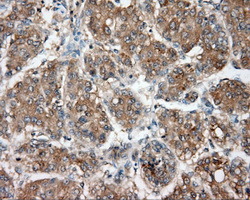
- Immunohistochemical staining of paraffin-embedded Carcinoma of liver tissue using anti-NIT2 mouse monoclonal antibody. (Heat-induced epitope retrieval by 10mM citric buffer, pH6.0, 100C for 10min, TA501109, Dilution 1:50)
- Validation comment
- IHC
- Submitted by
- OriGene (provider)
- Main image
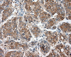
- Experimental details
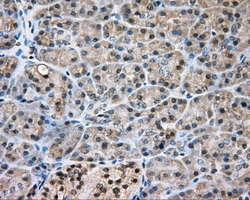
- Immunohistochemical staining of paraffin-embedded pancreas tissue within the normal limits using anti-NIT2 mouse monoclonal antibody. (Heat-induced epitope retrieval by 10mM citric buffer, pH6.0, 100C for 10min, TA501109, Dilution 1:50)
- Validation comment
- IHC

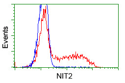
Supportive validation
- Submitted by
- OriGene (provider)
- Main image
- Experimental details
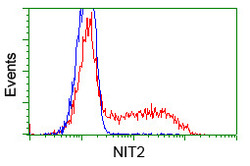
- HEK293T cells transfected with either pCMV6-ENTRY NIT2(RC210660)(Red) or empty vector control plasmid(Blue) were immunostained with anti-NIT2 mouse monoclonal(TA501109), and then analyzed by flow cytometry.
- Validation comment
- FC