 Explore
 Validate
 Learn
Learn14818-1-AP
antibody from Proteintech Group
Targeting: MICAL1
DKFZp434B1517, FLJ11937, FLJ21739, MICAL, NICAL
 Western blot
 ELISA
ELISAAntibody data
- Antibody Data
- Antigen structure
- References [15]
- Comments [0]
- Validations
- Western blot [1]
- Immunoprecipitation [1]
- Immunohistochemistry [2]
- Flow cytometry [1]
Submit
Validation data
Reference
Comment
Report error
- Product number
- 14818-1-AP - Provider product page

- Provider
- Proteintech Group
- Proper citation
- Proteintech Cat#14818-1-AP, RRID:AB_2143754
- Product name
- MICAL1 antibody
- Antibody type
- Polyclonal
- Description
- KD/KO validated MICAL1 antibody (Cat. #14818-1-AP) is a rabbit polyclonal antibody that shows reactivity with human, mouse and has been validated for the following applications: FC, IF, IHC, IP, WB,ELISA.
- Reactivity
- Human, Mouse
- Host
- Rabbit
- Conjugate
- Unconjugated
- Isotype
- IgG
- Vial size
- 20ul, 150ul
Submitted references Phosphorylation of MICAL2 by ARG promotes head and neck cancer tumorigenesis by regulating skeletal rearrangement.
MICAL1 regulates actin cytoskeleton organization, directional cell migration and the growth of human breast cancer cells as orthotopic xenograft tumours.
MICAL2 enhances branched actin network disassembly by oxidizing Arp3B-containing Arp2/3 complexes.
MICAL1 (molecule interacting with CasL 1) protects oligodendrocyte cells from oxidative injury through regulating apoptosis, autophagy in spinal cord injury.
TBC1D1 interacting proteins, VPS13A and VPS13C, regulate GLUT4 homeostasis in C2C12 myotubes.
F-actin disassembly factor MICAL1 binding to Myosin Va mediates cargo unloading during cytokinesis.
NEDD9 Facilitates Hypoxia-Induced Gastric Cancer Cell Migration via MICAL1 Related Rac1 Activation.
NEDD9 stimulated MMP9 secretion is required for invadopodia formation in oral squamous cell carcinoma.
MICAL1 facilitates breast cancer cell proliferation via ROS-sensitive ERK/cyclin D pathway.
Podocyte Shape Regulation by Semaphorin 3A and MICAL-1.
Amplification of F-Actin Disassembly and Cellular Repulsion by Growth Factor Signaling.
Oxidation of F-actin controls the terminal steps of cytokinesis.
MICAL1 controls cell invasive phenotype via regulating oxidative stress in breast cancer cells.
Semaphorin3a promotes advanced diabetic nephropathy.
Differential regulation of actin microfilaments by human MICAL proteins.
Zhang Z, Liu R, Wang Y, Wang Y, Shuai Y, Ke C, Jin R, Wang X, Luo J
Oncogene 2022 Jan;41(3):334-346
Oncogene 2022 Jan;41(3):334-346
MICAL1 regulates actin cytoskeleton organization, directional cell migration and the growth of human breast cancer cells as orthotopic xenograft tumours.
McGarry DJ, Armstrong G, Castino G, Mason S, Clark W, Shaw R, McGarry L, Blyth K, Olson MF
Cancer letters 2021 Oct 28;519:226-236
Cancer letters 2021 Oct 28;519:226-236
MICAL2 enhances branched actin network disassembly by oxidizing Arp3B-containing Arp2/3 complexes.
Galloni C, Carra D, Abella JVG, Kjær S, Singaravelu P, Barry DJ, Kogata N, Guérin C, Blanchoin L, Way M
The Journal of cell biology 2021 Aug 2;220(8)
The Journal of cell biology 2021 Aug 2;220(8)
MICAL1 (molecule interacting with CasL 1) protects oligodendrocyte cells from oxidative injury through regulating apoptosis, autophagy in spinal cord injury.
Xu C, Mao L, Tian H, Lin S, Zhao X, Lin J, Li D, Li X, Mei X
Neuroscience letters 2021 Apr 17;750:135712
Neuroscience letters 2021 Apr 17;750:135712
TBC1D1 interacting proteins, VPS13A and VPS13C, regulate GLUT4 homeostasis in C2C12 myotubes.
Hook SC, Chadt A, Heesom KJ, Kishida S, Al-Hasani H, Tavaré JM, Thomas EC
Scientific reports 2020 Oct 21;10(1):17953
Scientific reports 2020 Oct 21;10(1):17953
F-actin disassembly factor MICAL1 binding to Myosin Va mediates cargo unloading during cytokinesis.
Niu F, Sun K, Wei W, Yu C, Wei Z
Science advances 2020 Nov;6(45)
Science advances 2020 Nov;6(45)
NEDD9 Facilitates Hypoxia-Induced Gastric Cancer Cell Migration via MICAL1 Related Rac1 Activation.
Zhao S, Min P, Liu L, Zhang L, Zhang Y, Wang Y, Zhao X, Ma Y, Xie H, Zhu C, Jiang H, Du J, Gu L
Frontiers in pharmacology 2019;10:291
Frontiers in pharmacology 2019;10:291
NEDD9 stimulated MMP9 secretion is required for invadopodia formation in oral squamous cell carcinoma.
Grauzam S, Brock AM, Holmes CO, Tiedeken JA, Boniface SG, Pierson BN, Patterson DG, Coaxum SD, Neskey DM, Rosenzweig SA
Oncotarget 2018 May 22;9(39):25503-25516
Oncotarget 2018 May 22;9(39):25503-25516
MICAL1 facilitates breast cancer cell proliferation via ROS-sensitive ERK/cyclin D pathway.
Deng W, Wang Y, Zhao S, Zhang Y, Chen Y, Zhao X, Liu L, Sun S, Zhang L, Ye B, Du J
Journal of cellular and molecular medicine 2018 Jun;22(6):3108-3118
Journal of cellular and molecular medicine 2018 Jun;22(6):3108-3118
Podocyte Shape Regulation by Semaphorin 3A and MICAL-1.
Tufro A
Methods in molecular biology (Clifton, N.J.) 2017;1493:393-399
Methods in molecular biology (Clifton, N.J.) 2017;1493:393-399
Amplification of F-Actin Disassembly and Cellular Repulsion by Growth Factor Signaling.
Yoon J, Kim SB, Ahmed G, Shay JW, Terman JR
Developmental cell 2017 Jul 24;42(2):117-129.e8
Developmental cell 2017 Jul 24;42(2):117-129.e8
Oxidation of F-actin controls the terminal steps of cytokinesis.
Frémont S, Hammich H, Bai J, Wioland H, Klinkert K, Rocancourt M, Kikuti C, Stroebel D, Romet-Lemonne G, Pylypenko O, Houdusse A, Echard A
Nature communications 2017 Feb 23;8:14528
Nature communications 2017 Feb 23;8:14528
MICAL1 controls cell invasive phenotype via regulating oxidative stress in breast cancer cells.
Deng W, Wang Y, Gu L, Duan B, Cui J, Zhang Y, Chen Y, Sun S, Dong J, Du J
BMC cancer 2016 Jul 18;16:489
BMC cancer 2016 Jul 18;16:489
Semaphorin3a promotes advanced diabetic nephropathy.
Aggarwal PK, Veron D, Thomas DB, Siegel D, Moeckel G, Kashgarian M, Tufro A
Diabetes 2015 May;64(5):1743-59
Diabetes 2015 May;64(5):1743-59
Differential regulation of actin microfilaments by human MICAL proteins.
Giridharan SS, Rohn JL, Naslavsky N, Caplan S
Journal of cell science 2012 Feb 1;125(Pt 3):614-24
Journal of cell science 2012 Feb 1;125(Pt 3):614-24
No comments: Submit comment
Supportive validation
- Submitted by
- Proteintech Group (provider)
- Main image

- Experimental details
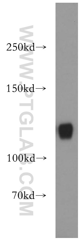
- Jurkat cells were subjected to SDS PAGE followed by western blot with 14818-1-AP(MICAL1 antibody) at dilution of 1:1000
- Sample type
- cell line
Supportive validation
- Submitted by
- Proteintech Group (provider)
- Main image
- Experimental details
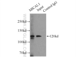
- IP Result of anti-MICAL1 (IP:14818-1-AP, 5ug; Detection:14818-1-AP 1:1000) with HeLa cells lysate 2000ug.
- Sample type
- cell line

Supportive validation
- Submitted by
- Proteintech Group (provider)
- Main image

- Experimental details
- Immunohistochemical of paraffin-embedded human lung using 14818-1-AP(MICAL1 antibody) at dilution of 1:100 (under 40x lens)
- Sample type
- tissue

- Submitted by
- Proteintech Group (provider)
- Main image

- Experimental details
- Immunohistochemical of paraffin-embedded human lung using 14818-1-AP(MICAL1 antibody) at dilution of 1:100 (under 10x lens)
- Sample type
- tissue

Supportive validation
- Submitted by
- Proteintech Group (provider)
- Main image
- Experimental details
- The MICAL1 antibody from Proteintech is a rabbit polyclonal antibody to a recombinant protein of human MICAL1. This antibody recognizes human, mouse antigen. The MICAL1 antibody has been validated for the following applications: ELISA, WB, IHC, IP, FC analysis.
