Explore
 Validate
 Learn
Learn Western blot
Antibody data
- Antibody Data
- Antigen structure
- References [1]
- Comments [0]
- Validations
- Western blot [6]
- Immunocytochemistry [1]
- Immunohistochemistry [2]
Submit
Validation data
Reference
Comment
Report error
- Product number
- GTX111664 - Provider product page

- Provider
- GeneTex
- Proper citation
- GeneTex Cat#GTX111664, RRID:AB_1950539
- Product name
- IDE antibody [N3C1], Internal
- Antibody type
- Polyclonal
- Reactivity
- Human, Mouse, Rat
- Host
- Rabbit
Submitted references Mice lacking the transcriptional regulator Bhlhe40 have enhanced neuronal excitability and impaired synaptic plasticity in the hippocampus.
Hamilton KA, Wang Y, Raefsky SM, Berkowitz S, Spangler R, Suire CN, Camandola S, Lipsky RH, Mattson MP
PloS one 2018;13(5):e0196223
PloS one 2018;13(5):e0196223
No comments: Submit comment
Supportive validation
- Submitted by
- GeneTex (provider)
- Main image
- Experimental details
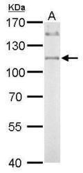
- IDE antibody [N3C1], Internal detects IDE protein by western blot analysis.A. 50 ?g mouse liver lysate/extract 7.5% SDS-PAGEIDE antibody [N3C1], Internal (GTX111664) dilution: 1:1000 The HRP-conjugated anti-rabbit IgG antibody (GTX213110-01) was used to detect the primary antibody.

- Submitted by
- GeneTex (provider)
- Main image
- Experimental details
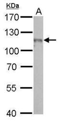
- IDE antibody [N3C1], Internal detects IDE protein by western blot analysis.A. 50 ?g rat brain lysate/extract7.5% SDS-PAGEIDE antibody [N3C1], Internal (GTX111664) dilution: 1:500 The HRP-conjugated anti-rabbit IgG antibody (GTX213110-01) was used to detect the primary antibody.

- Submitted by
- GeneTex (provider)
- Main image
- Experimental details
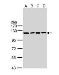
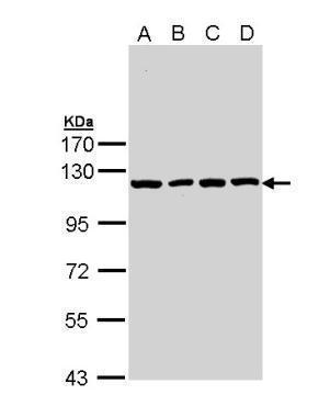
- Sample (30 ?g of whole cell lysate) A: A431 (GTX27909) B: H1299 C: HeLa D: HepG2 (GTX27900) 7.5% SDS PAGE GTX111664 diluted at 1:10000 The HRP-conjugated anti-rabbit IgG antibody (GTX213110-01) was used to detect the primary antibody.
- Submitted by
- GeneTex (provider)
- Main image

- Experimental details
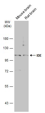
- Various tissue extracts (50 ?g) were separated by 7.5% SDS-PAGE, and the membrane was blotted with IDE antibody [N3C1], Internal (GTX111664) diluted at 1:1000. The HRP-conjugated anti-rabbit IgG antibody (GTX213110-01) was used to detect the primary antibody.
- Submitted by
- GeneTex (provider)
- Main image
- Experimental details
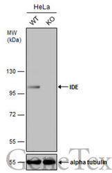
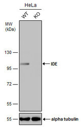
- Wild-type (WT) and IDE knockout (KO) HeLa cell extracts (30 ?g) were separated by 7.5% SDS-PAGE, and the membrane was blotted with IDE antibody [N3C1], Internal (GTX111664) diluted at 1:2500. The HRP-conjugated anti-rabbit IgG antibody (GTX213110-01) was used to detect the primary antibody.
- Submitted by
- GeneTex (provider)
- Main image
- Experimental details
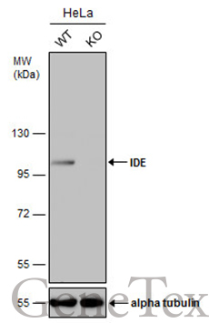
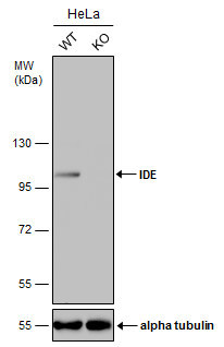
- Wild-type (WT) and IDE knockout (KO) HeLa cell extracts (30 ?g) were separated by 7.5% SDS-PAGE, and the membrane was blotted with IDE antibody [N3C1], Internal (GTX111664) diluted at 1:2500. The HRP-conjugated anti-rabbit IgG antibody (GTX213110-01) was used to detect the primary antibody.
Supportive validation
- Submitted by
- GeneTex (provider)
- Main image
- Experimental details
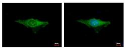
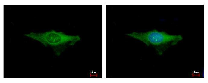
- IDE antibody [N3C1], Internal detects IDE protein at cytoplasm by immunofluorescent analysis. Sample: HeLa cells were fixed in ice-cold MeOH for 5 min.Green: IDE protein stained by IDE antibody [N3C1], Internal (GTX111664) diluted at 1:500.Blue: Hoechst 33343 staining.
Supportive validation
- Submitted by
- GeneTex (provider)
- Main image
- Experimental details
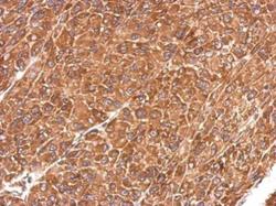
- Immunohistochemical analysis of paraffin-embedded U87 xenograft, using insulin degrading enzyme(GTX111664) antibody at 1:500 dilution.

- Submitted by
- GeneTex (provider)
- Main image
- Experimental details
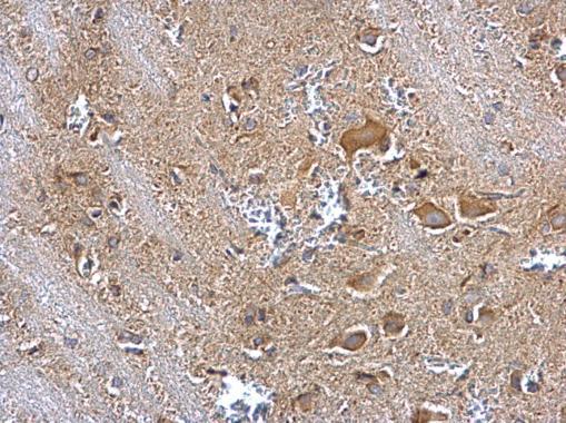
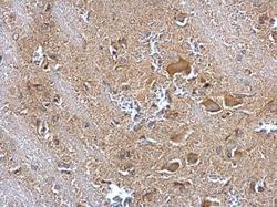
- IDE antibody [N3C1], Internal detects IDE protein at cytosol on rat brain stem by immunohistochemical analysis. Sample: Paraffin-embedded rat brain stem. IDE antibody [N3C1], Internal (GTX111664) dilution: 1:500.