 Explore
 Validate
 Learn
Learn Western blot
 Immunohistochemistry
Antibody data
- Antibody Data
- Antigen structure
- References [7]
- Comments [0]
- Validations
- Immunohistochemistry [1]
- Flow cytometry [1]
Submit
Validation data
Reference
Comment
Report error
- Product number
- AF619 - Provider product page

- Provider
- R&D Systems
- Product name
- Human Tie-1 Antibody
- Antibody type
- Polyclonal
- Description
- Antigen Affinity-purified. Detects human Tie-1 in direct ELISAs and Western blots. In direct ELISAs, approximately 50% cross-reactivity with recombinant mouse Tie-1 is observed and less than 1% cross-reactivity with recombinant human Tie-2 is observed.
- Reactivity
- Human
- Host
- Goat
- Conjugate
- Unconjugated
- Antigen sequence
P35590- Isotype
- IgG
- Vial size
- 100 ug
- Concentration
- LYOPH
- Storage
- Use a manual defrost freezer and avoid repeated freeze-thaw cycles. 12 months from date of receipt, -20 to -70 °C as supplied. 1 month, 2 to 8 °C under sterile conditions after reconstitution. 6 months, -20 to -70 °C under sterile conditions after reconstitution.
Submitted references Structural basis of Tie2 activation and Tie2/Tie1 heterodimerization.
Polydom Is an Extracellular Matrix Protein Involved in Lymphatic Vessel Remodeling.
Endothelial destabilization by angiopoietin-2 via integrin β1 activation.
Regulated proteolytic processing of Tie1 modulates ligand responsiveness of the receptor-tyrosine kinase Tie2.
Interaction between Tie receptors modulates angiogenic activity of angiopoietin2 in endothelial progenitor cells.
Expansion of human SCID-repopulating cells under hypoxic conditions.
Evidence for heterotypic interaction between the receptor tyrosine kinases TIE-1 and TIE-2.
Leppänen VM, Saharinen P, Alitalo K
Proceedings of the National Academy of Sciences of the United States of America 2017 Apr 25;114(17):4376-4381
Proceedings of the National Academy of Sciences of the United States of America 2017 Apr 25;114(17):4376-4381
Polydom Is an Extracellular Matrix Protein Involved in Lymphatic Vessel Remodeling.
Morooka N, Futaki S, Sato-Nishiuchi R, Nishino M, Totani Y, Shimono C, Nakano I, Nakajima H, Mochizuki N, Sekiguchi K
Circulation research 2017 Apr 14;120(8):1276-1288
Circulation research 2017 Apr 14;120(8):1276-1288
Endothelial destabilization by angiopoietin-2 via integrin β1 activation.
Hakanpaa L, Sipila T, Leppanen VM, Gautam P, Nurmi H, Jacquemet G, Eklund L, Ivaska J, Alitalo K, Saharinen P
Nature communications 2015 Jan 30;6:5962
Nature communications 2015 Jan 30;6:5962
Regulated proteolytic processing of Tie1 modulates ligand responsiveness of the receptor-tyrosine kinase Tie2.
Marron MB, Singh H, Tahir TA, Kavumkal J, Kim HZ, Koh GY, Brindle NP
The Journal of biological chemistry 2007 Oct 19;282(42):30509-17
The Journal of biological chemistry 2007 Oct 19;282(42):30509-17
Interaction between Tie receptors modulates angiogenic activity of angiopoietin2 in endothelial progenitor cells.
Kim KL, Shin IS, Kim JM, Choi JH, Byun J, Jeon ES, Suh W, Kim DK
Cardiovascular research 2006 Dec 1;72(3):394-402
Cardiovascular research 2006 Dec 1;72(3):394-402
Expansion of human SCID-repopulating cells under hypoxic conditions.
Danet GH, Pan Y, Luongo JL, Bonnet DA, Simon MC
The Journal of clinical investigation 2003 Jul;112(1):126-35
The Journal of clinical investigation 2003 Jul;112(1):126-35
Evidence for heterotypic interaction between the receptor tyrosine kinases TIE-1 and TIE-2.
Marron MB, Hughes DP, Edge MD, Forder CL, Brindle NP
The Journal of biological chemistry 2000 Dec 15;275(50):39741-6
The Journal of biological chemistry 2000 Dec 15;275(50):39741-6
No comments: Submit comment
Supportive validation
- Submitted by
- R&D Systems (provider)
- Main image
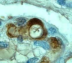
- Experimental details
- Tie-1 in Human Placenta. Tie-1 was detected in immersion fixed paraffin-embedded sections of human placenta using 8 µg/mL Goat Anti-Human Tie-1 Antigen Affinity-purified Polyclonal Antibody (Catalog # AF619) overnight at 4 °C. Tissue was stained with the Anti-Goat HRP-DAB Cell & Tissue Staining Kit (brown; Catalog # CTS008) and counter-stained with hematoxylin (blue). View our protocol for Chromogenic IHC Staining of Paraffin-embedded Tissue Sections.

Supportive validation
- Submitted by
- R&D Systems (provider)
- Main image
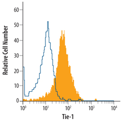
- Experimental details
- Detection of Tie-1 in HUVEC Human Cells by Flow Cytometry. HUVEC human umbilical vein endothelial cells were stained with Goat Anti-Human Tie-1 Antigen Affinity-purified Polyclonal Antibody (Catalog # AF619, filled histogram) or control antibody (Catalog # AB-108-C, open histogram), followed by Phycoerythrin-conjugated Anti-Goat IgG Secondary Antibody (Catalog # F0107).
