Explore
 Validate
 Learn
Learn Western blot
 Immunocytochemistry
Antibody data
- Antibody Data
- Antigen structure
- References [2]
- Comments [0]
- Validations
- Western blot [3]
Submit
Validation data
Reference
Comment
Report error
- Product number
- MA1-22151 - Provider product page

- Provider
- Invitrogen Antibodies
- Product name
- Anti-beta COP Monoclonal Antibody (maD)
- Antibody type
- Monoclonal
- Antigen
- Synthetic peptide
- Description
- Recommended positive controls: CHO, stacked Golgi membranes from rat liver. In immunofluorescence, use methanol/acetone or paraformaldehyde fixation, followed by a permeabilization step. Strong immunofluorescent staining is confined mainly to the central perinuclear area in the cell.
- Reactivity
- Human, Mouse, Rat, Hamster
- Host
- Mouse
- Isotype
- IgG
- Antibody clone number
- maD
- Vial size
- 100 µL
- Concentration
- Conc. Not Determined
- Storage
- Store at 4°C short term. For long term storage, store at -20°C, avoiding freeze/thaw cycles.
Submitted references Golgi fragmentation is Rab and SNARE dependent in cellular models of Parkinson's disease.
Bidirectional transport by distinct populations of COPI-coated vesicles.
Rendón WO, Martínez-Alonso E, Tomás M, Martínez-Martínez N, Martínez-Menárguez JA
Histochemistry and cell biology 2013 May;139(5):671-84
Histochemistry and cell biology 2013 May;139(5):671-84
Bidirectional transport by distinct populations of COPI-coated vesicles.
Orci L, Stamnes M, Ravazzola M, Amherdt M, Perrelet A, Söllner TH, Rothman JE
Cell 1997 Jul 25;90(2):335-49
Cell 1997 Jul 25;90(2):335-49
No comments: Submit comment
Supportive validation
- Submitted by
- Invitrogen Antibodies (provider)
- Main image
- Experimental details
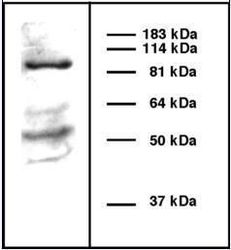
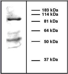
- Western blot analysis of beta COP using a beta COP monoclonal antibody (Product # MA1-22151).
- Submitted by
- Invitrogen Antibodies (provider)
- Main image
- Experimental details
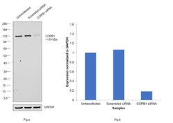
- Knockdown of COPB1 was achieved by transfecting HeLa with COPB1 specific siRNAs (Silencer® select Product # s3372). Western blot analysis (Fig. a) was performed using membrane enriched extracts from the COPB1 knockdown cells (lane 3), non-specific scrambled siRNA transfected cells (lane 2) and untransfected cells (lane 1). The blot was probed with COP Monoclonal Antibody (maD) (Product # MA1-22151, 1:1000 dilution) and Goat anti-Mouse IgG (H+L) Superclonal™ Recombinant Secondary Antibody, HRP (Product # A28177, 1:4000 dilution). Densitometric analysis of this western blot is shown in histogram (Fig. b). Decrease in signal upon siRNA mediated knock down confirms that antibody is specific to COPB1.

- Submitted by
- Invitrogen Antibodies (provider)
- Main image
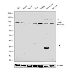
- Experimental details
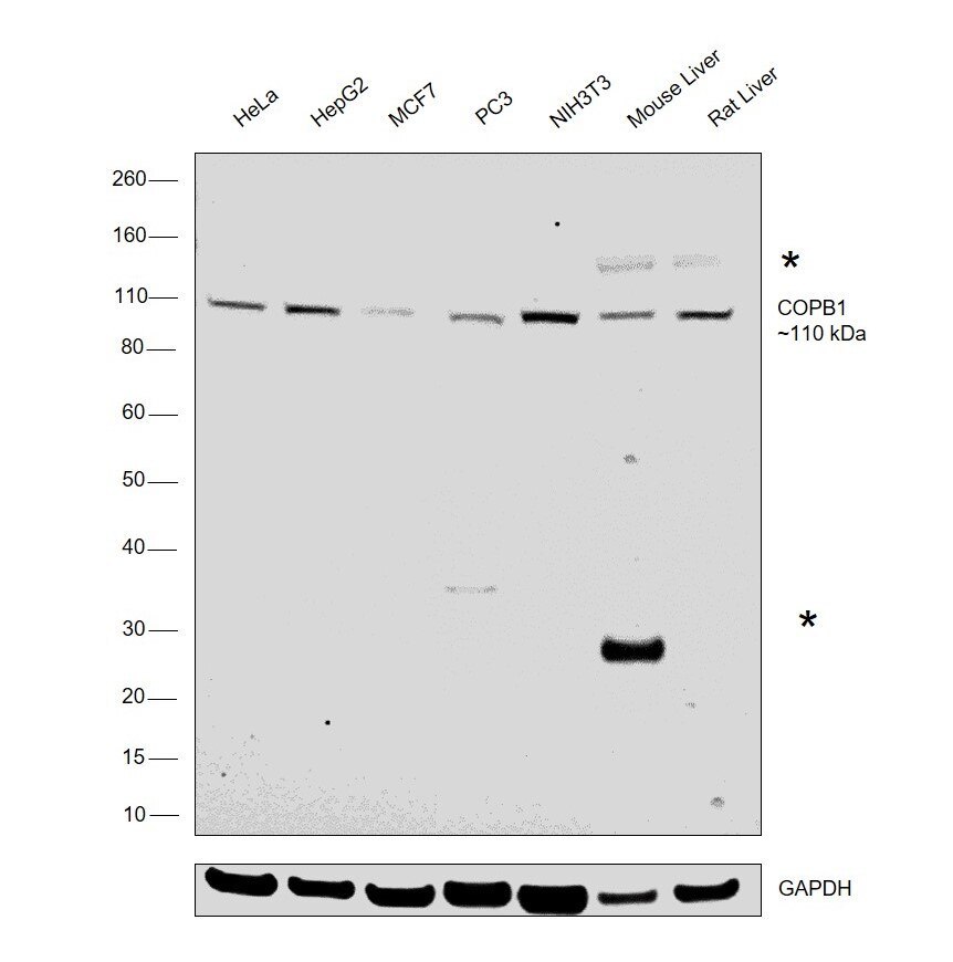
- Western blot was performed using beta COP Monoclonal Antibody (maD) (Product # MA1-22151) and a 110 kDa band corresponding to COPB1 was observed across cell lines and tissue extracts along with an uncharacterized band (*) at ~130 kDa in the tissues tested. Also, an additional band (*) at ~25 kDa corresponding to circulating tissue IgG was observed in mouse tissue. Membrane enriched extracts (30 µg lysate) of HeLa (Lane 1), HepG2 (Lane 2), MCF7 (Lane 3), PC3 (Lane 4), NIH3T3 (Lane 5), tissue extracts (30ug lysate) of Mouse Liver (Lane 6) and Rat Liver (Lane 7) were electrophoresed using NuPAGE® 4-12 % Bis-Tris gel (Product # NP0322BOX). Resolved proteins were then transferred onto a nitrocellulose membrane (Product # IB23001) by iBlot® 2 Dry Blotting System (Product # IB21001).The blot was probed with the primary antibody (1:1000 dilution) and detected by chemiluminescence with Goat anti-Mouse IgG (H+L), Superclonal™ Recombinant Secondary Antibody, HRP (Product # A28177, 1:4000 dilution) using the iBright FL 1000 (Product # A32752). Chemiluminescent detection was performed using Novex® ECL Chemiluminescent Substrate Reagent Kit (Product # WP20005).