Explore
 Validate
 Learn
Learn Western blot
 Immunoprecipitation
Antibody data
- Antibody Data
- Antigen structure
- References [0]
- Comments [0]
- Validations
- Western blot [6]
- Immunohistochemistry [4]
- Chromatin Immunoprecipitation [1]
- Other assay [1]
Submit
Validation data
Reference
Comment
Report error
- Product number
- A300-815A - Provider product page

- Provider
- Invitrogen Antibodies
- Product name
- TIF1 Alpha/TRIM24 Polyclonal Antibody
- Antibody type
- Polyclonal
- Antigen
- Other
- Reactivity
- Human, Mouse
- Host
- Rabbit
- Isotype
- IgG
- Vial size
- 100 µL
- Concentration
- 0.20 mg/mL
- Storage
- 4° C
No comments: Submit comment
Supportive validation
- Submitted by
- Invitrogen Antibodies (provider)
- Main image

- Experimental details
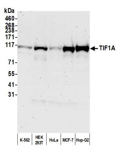
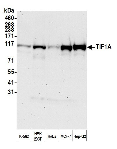
- Detection of human TIF1A by western blot. Samples: Whole cell lysate (50 µg) from K-562, HEK293T, HeLa, MCF-7, and Hep-G2 cells prepared using NETN lysis buffer. Antibody: Affinity purified rabbit anti-TIF1A antibody (Product # A300-815A lot 3) used for WB at 0.1 µg/mL. Detection: Chemiluminescence with an exposure time of 75 seconds.
- Submitted by
- Invitrogen Antibodies (provider)
- Main image

- Experimental details
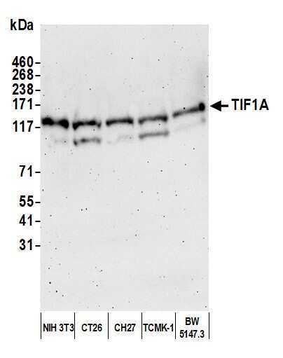
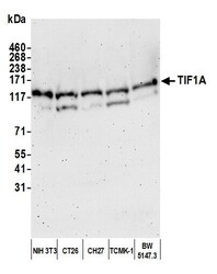
- Detection of mouse TIF1A by western blot. Samples: Whole cell lysate (50 µg) from NIH 3T3, CT26, CH27, TCMK-1, and BW5147.3 cells prepared using NETN lysis buffer. Antibody: Affinity purified rabbit anti-TIF1A antibody (Product # A300-815A lot 3) used for WB at 0.1 µg/mL. Detection: Chemiluminescence with an exposure time of 3 minutes.
- Submitted by
- Invitrogen Antibodies (provider)
- Main image
- Experimental details
- Detection of human TIF1A by western blot. Samples: Whole cell lysate (50 µg) from K-562, HEK293T, HeLa, MCF-7, and Hep-G2 cells prepared using NETN lysis buffer. Antibody: Affinity purified rabbit anti-TIF1A antibody (Product # A300-815A lot 3) used for WB at 0.1 µg/mL. Detection: Chemiluminescence with an exposure time of 75 seconds.
- Submitted by
- Invitrogen Antibodies (provider)
- Main image
- Experimental details
- Detection of mouse TIF1A by western blot. Samples: Whole cell lysate (50 µg) from NIH 3T3, CT26, CH27, TCMK-1, and BW5147.3 cells prepared using NETN lysis buffer. Antibody: Affinity purified rabbit anti-TIF1A antibody (Product # A300-815A lot 3) used for WB at 0.1 µg/mL. Detection: Chemiluminescence with an exposure time of 3 minutes.

- Submitted by
- Invitrogen Antibodies (provider)
- Main image

- Experimental details
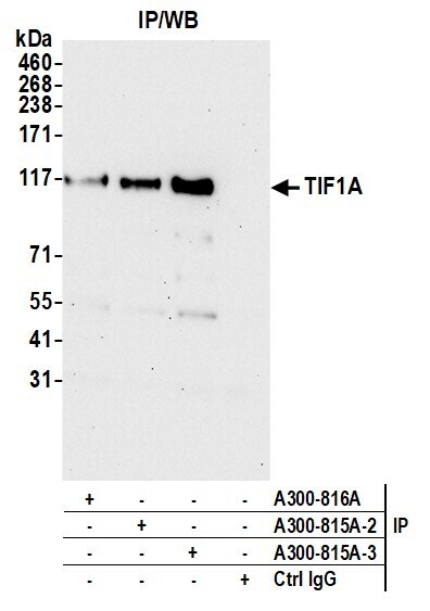
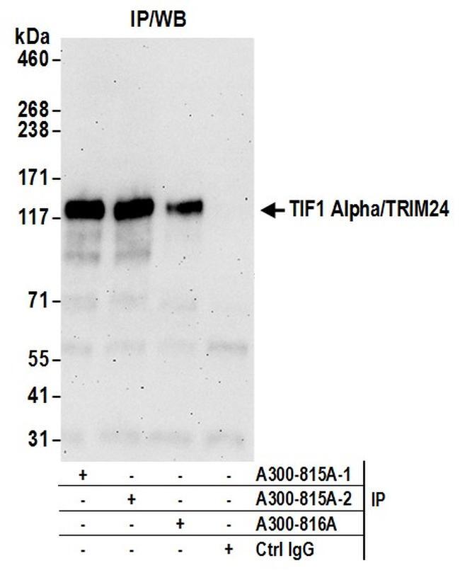
- Detection of human TIF1 Alpha/TRIM24 by western blot of immunoprecipitates. Samples: Whole cell lysate (0.5 or 1.0 mg per IP reaction; 20% of IP loaded) from HeLa cells prepared using NETN lysis buffer. Antibodies: Affinity purified rabbit anti-TIF1 Alpha/TRIM24 antibody A300-815A (lot A300-815A-2) used for IP at 6 µg per reaction. TIF1 Alpha/TRIM24 was also immunoprecipitated by a previous lot of this antibody (lot A300-815A-1) and rabbit anti-TIF1 Alpha/TRIM24 antibody A300-816A For blotting immunoprecipitated TIF1 Alpha/TRIM24, A300-815A was used at 1 µg/ml. Detection: Chemiluminescence with an exposure time of 3 minutes.
- Submitted by
- Invitrogen Antibodies (provider)
- Main image
- Experimental details
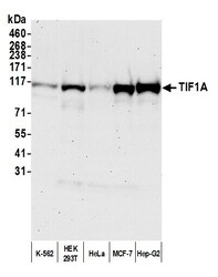
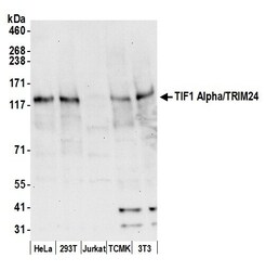
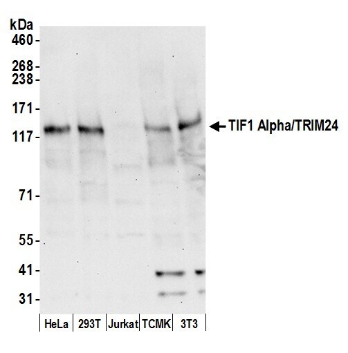
- Detection of human and mouse TIF1 Alpha/TRIM24 by western blot. Samples: Whole cell lysate (50 µg) from HeLa, 293T, Jurkat, mouse TCMK-1, and mouse NIH3T3 cells prepared using NETN lysis buffer. Antibody: Affinity purified rabbit anti-TIF1 Alpha/TRIM24 antibody A300-815A (lot A300-815A-2) used for WB at 0.1 µg/ml. Detection: Chemiluminescence with an exposure time of 30 seconds.
Supportive validation
- Submitted by
- Invitrogen Antibodies (provider)
- Main image
- Experimental details
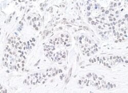
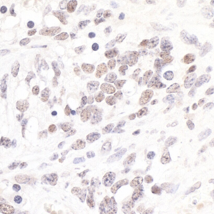
- Detection of human TIF1 alpha/TRIM24 by immunohistochemistry. Sample: FFPE section of human breast carcinoma. Antibody: Affinity purified rabbit anti-TIF1 alpha/TRIM24 (Product # A300-815A Lot 3) used at a dilution of 1:200 (1 µg/mL). Detection: DAB.

- Submitted by
- Invitrogen Antibodies (provider)
- Main image
- Experimental details
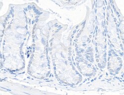
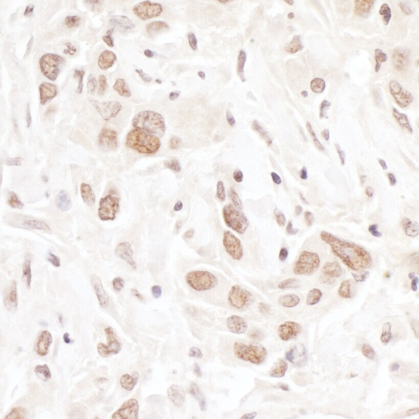
- Detection of mouse TIF1 alpha/TRIM24 by immunohistochemistry. Sample: FFPE section of mouse intestine. Antibody: Affinity purified rabbit anti-TIF1 alpha/TRIM24 (Product # A300-815A Lot 3) used at a dilution of 1:200 (1 µg/mL). Detection: DAB.

- Submitted by
- Invitrogen Antibodies (provider)
- Main image
- Experimental details
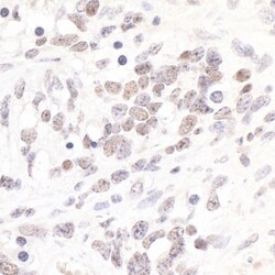
- Detection of mouse TIF1 alpha/TRIM24 by immunohistochemistry. Sample: FFPE section of mouse teratoma. Antibody: Affinity purified rabbit anti-TIF1 alpha/TRIM24 (Cat. No. A300-815A Lot2) used at a dilution of 1:200 (1µg/ml). Detection: DAB.
- Submitted by
- Invitrogen Antibodies (provider)
- Main image

- Experimental details
- Detection of human TIF1 alpha/TRIM24 by immunohistochemistry. Sample: FFPE section of human breast carcinoma. Antibody: Affinity purified rabbit anti- TIF1 alpha/TRIM24 (Cat. No. A300-815A Lot2) used at a dilution of 1:200 (1µg/ml). Detection: DAB.
Supportive validation
- Submitted by
- Invitrogen Antibodies (provider)
- Main image

- Experimental details
- Localization of TIF1 Alpha/TRIM24 Binding Sites by ChIP-sequencing.Chromatin from Breast cancer cell line T47D was immunoprecipitated with anti-TIF1 Alpha/TRIM24 antibody A300-815A and analyzed by DNA sequencing. The figure illustrates the peak distribution of TIF1 Alpha/TRIM24 binding within a 250 Kb region of chromosome 19 as detected using anti-TIF1 Alpha/TRIM24 antibody A300-815A. ChIP-seq validation performed by Active Motif, Carlsbad, CA.

Supportive validation
- Submitted by
- Invitrogen Antibodies (provider)
- Main image

- Experimental details
- Detection of human TIF1A by western blot of immunoprecipitates. Samples: Whole cell lysate (1.0 mg per IP reaction; 20% of IP loaded) from HEK293T cells prepared using NETN lysis buffer. Antibodies: Affinity purified rabbit anti-TIF1A antibody (Product # A300-815A lot 3) used for IP at 6 µg per reaction. TIF1A was also immunoprecipitated by a previous lot of this antibody (Product # A300-815A lot 2) and a second antibody against a different epitope of TIF1A (Product # A300-816A). For blotting immunoprecipitated TIF1A (Product # A300-815A) was used at 0.04 µg/mL. Detection: Chemiluminescence with an exposure time of 30 seconds.