 Explore
 Validate
 Learn
Learn Western blot
 Immunocytochemistry
Antibody data
- Antibody Data
- Antigen structure
- References [0]
- Comments [0]
- Validations
- Western blot [3]
- Immunocytochemistry [3]
- Immunohistochemistry [1]
Submit
Validation data
Reference
Comment
Report error
- Product number
- HPA059070 - Provider product page

- Provider
- Atlas Antibodies
- Proper citation
- Atlas Antibodies Cat#HPA059070, RRID:AB_2683896
- Product name
- Anti-PRPF19
- Antibody type
- Polyclonal
- Reactivity
- Human
- Host
- Rabbit
- Conjugate
- Unconjugated
- Antigen sequence
LQDKATVLTTERKKRGKTVPEELVKPEELSKYRQV
ASHVGLHSASIPGILALDLCPSDTNKILTGGADKN
VVVFDKSSEQILATLKGHTKKVTSV- Isotype
- IgG
- Vial size
- 100 µl
- Storage
- Store at +4°C for short term storage. Long time storage is recommended at -20°C.
No comments: Submit comment
Supportive validation
Supportive validation
- Submitted by
- Atlas Antibodies (provider)
- Enhanced method
- Genetic validation
- Main image

- Experimental details
- Western blot analysis in U2OS cells transfected with control siRNA, target specific siRNA probe #1 and #2, using Anti-PRPF19 antibody. Remaining relative intensity is presented.

- Submitted by
- Atlas Antibodies (provider)
- Enhanced method
- Independent antibody validation
- Main image

- Experimental details
- Western blot analysis using Anti-PRPF19 antibody HPA059070 (A) shows similar pattern to independent antibody HPA038051 (B).

Supportive validation
- Submitted by
- Atlas Antibodies (provider)
- Main image
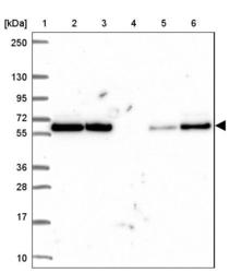
- Experimental details
- Lane 1: Marker [kDa] 250, 130, 95, 72, 55, 36, 28, 17, 10Lane 2: Human cell line RT-4Lane 3: Human cell line U-251 MGLane 4: Human plasmaLane 5: Human Liver tissueLane 6: Human Tonsil tissue

Enhanced validation
Supportive validation
- Submitted by
- 55af80e3e0991
- Enhanced method
- Genetic validation
- Main image

- Experimental details
- Confocal images of immunofluorescently stained human U-2 OS cells.The protein PRPF19 is shown in green and the microtubules in red. The image to the left show cells transfected with control siRNA and the image to the right show cells where PRPF19 has been downregulated with specific siRNA.
- Sample type
- U-2 OS cells
- Primary Ab dilution
- 1:58
- Secondary Ab
- Secondary Ab
- Secondary Ab dilution
- 1:800
- Knockdown/Genetic Approaches Application
- Immunocytochemistry

Supportive validation
- Submitted by
- Atlas Antibodies (provider)
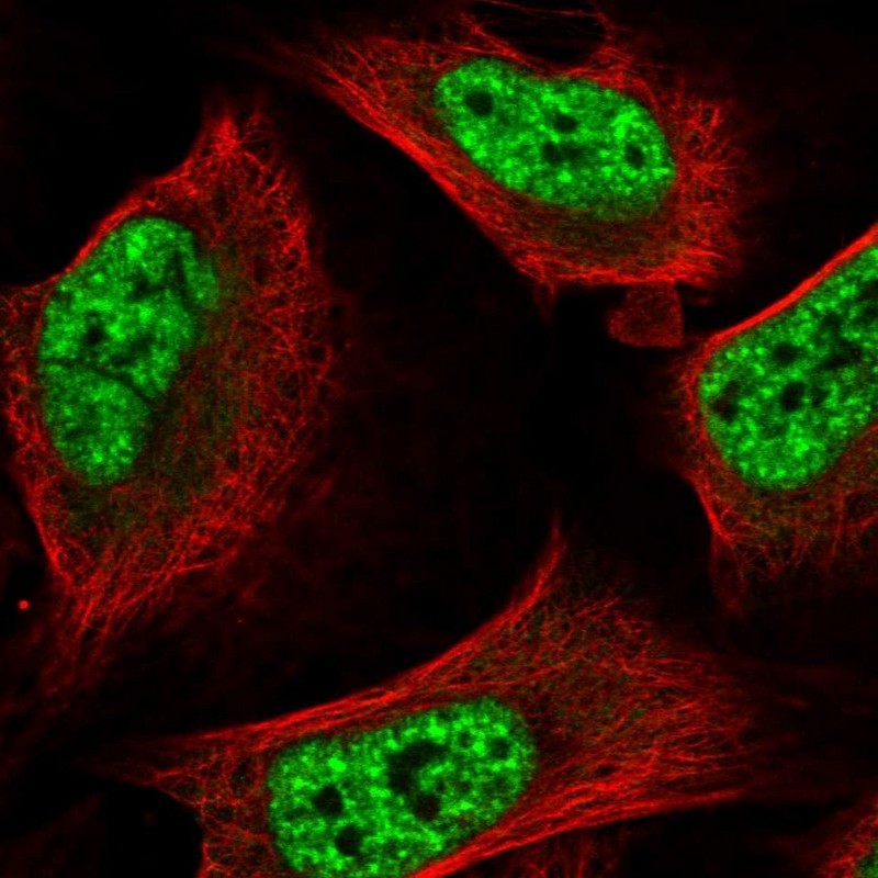
- Main image

- Experimental details
- Immunofluorescent staining of human cell line U-2 OS shows localization to nuclear speckles.
- Submitted by
- Atlas Antibodies (provider)
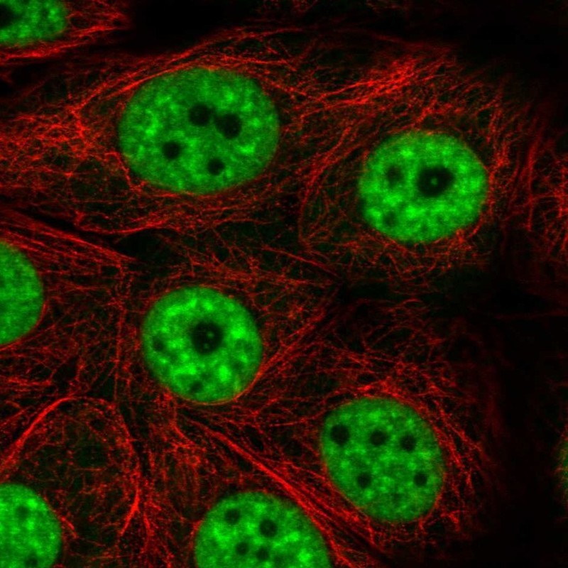
- Main image

- Experimental details
- Immunofluorescent staining of human cell line MCF7 shows localization to nuclear speckles.
- Sample type
- HUMAN
Supportive validation
- Submitted by
- Atlas Antibodies (provider)
- Main image

- Experimental details
- Immunohistochemical staining of human kidney shows strong nuclear positivity in cells in renal tubules and cells in glomeruli.
- Sample type
- HUMAN
