 Explore
 Validate
 Learn
LearnGTX101468
antibody from GeneTex
Targeting: P4HB
DSI, ERBA2L, GIT, P4Hbeta, PDI, PDIA1, PO4DB, PO4HB, PROHB
 Western blot
 Immunoelectron microscopy
Antibody data
- Antibody Data
- Antigen structure
- References [4]
- Comments [0]
- Validations
- Western blot [2]
- Immunocytochemistry [2]
- Immunohistochemistry [2]
Submit
Validation data
Reference
Comment
Report error
- Product number
- GTX101468 - Provider product page

- Provider
- GeneTex
- Proper citation
- GeneTex Cat#GTX101468, RRID:AB_1241147
- Product name
- PDI antibody [N1N3]
- Antibody type
- Polyclonal
- Reactivity
- Human, Mouse
- Host
- Rabbit
Submitted references Downregulation of proteins involved in the endoplasmic reticulum stress response and Nrf2-ARE signaling in lymphoblastoid cells of spinocerebellar ataxia type 17.
High glucose-induced proteome alterations in hepatocytes and its possible relevance to diabetic liver disease.
PI(4,5)P(2)-dependent and Ca(2+)-regulated ER-PM interactions mediated by the extended synaptotagmins.
Transcriptional activation of endoplasmic reticulum chaperone GRP78 by HCMV IE1-72 protein.
Lee LC, Weng YT, Wu YR, Soong BW, Tseng YC, Chen CM, Lee-Chen GJ
Journal of neural transmission (Vienna, Austria : 1996) 2014 Jun;121(6):601-10
Journal of neural transmission (Vienna, Austria : 1996) 2014 Jun;121(6):601-10
High glucose-induced proteome alterations in hepatocytes and its possible relevance to diabetic liver disease.
Chen JY, Chou HC, Chen YH, Chan HL
The Journal of nutritional biochemistry 2013 Nov;24(11):1889-910
The Journal of nutritional biochemistry 2013 Nov;24(11):1889-910
PI(4,5)P(2)-dependent and Ca(2+)-regulated ER-PM interactions mediated by the extended synaptotagmins.
Giordano F, Saheki Y, Idevall-Hagren O, Colombo SF, Pirruccello M, Milosevic I, Gracheva EO, Bagriantsev SN, Borgese N, De Camilli P
Cell 2013 Jun 20;153(7):1494-509
Cell 2013 Jun 20;153(7):1494-509
Transcriptional activation of endoplasmic reticulum chaperone GRP78 by HCMV IE1-72 protein.
Shi-Chen Ou D, Lee SB, Chu CS, Chang LH, Chung BC, Juan LJ
Cell research 2011 Apr;21(4):642-53
Cell research 2011 Apr;21(4):642-53
No comments: Submit comment
Supportive validation
- Submitted by
- GeneTex (provider)
- Main image

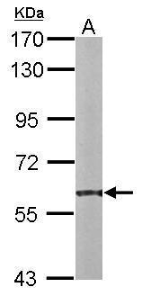
- Experimental details
- Sample (20 ug of whole cell lysate) A: mouse liver 7.5% SDS PAGE GTX101468 diluted at 1:10000
- Submitted by
- GeneTex (provider)
- Main image
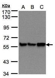
- Experimental details
- Sample(30 £gg of whole cell lysate)A:293TB:A431(GTX27909)C:H12997.5% SDS PAGEGTX101468 diluted at 1:500

Supportive validation
- Submitted by
- GeneTex (provider)
- Main image
- Experimental details
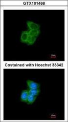
- Immunofluorescence analysis of methanol-fixed A431, using PDI(GTX101468) antibody at 1:200 dilution.

- Submitted by
- GeneTex (provider)
- Main image

- Experimental details
- PDI antibody [N1N3] detects PDI protein at endoplasmic reticulum by immunofluorescent analysis.Sample: HeLa cells were fixed in 4% paraformaldehyde at RT for 15 min.Green: PDI protein stained by PDI antibody [N1N3] (GTX101468) diluted at 1:100.Red: phalloidin, a cytoskeleton marker, stained by phalloidin (invitrogen, A12380) diluted at 1:200.Blue: Hoechst 33342 staining.

Supportive validation
- Submitted by
- GeneTex (provider)
- Main image

- Experimental details
- Immunohistochemical analysis of paraffin-embedded H520 xenograft, using PDI(GTX101468) antibody at 1:100 dilution.

- Submitted by
- GeneTex (provider)
- Main image

- Experimental details
- PDI antibody [N1N3] detects PDI protein at cytosol and membrane on mouse colon by immunohistochemical analysis. Sample: Paraffin-embedded mouse colon. PDI antibody [N1N3] (GTX101468) dilution: 1:500.
