Explore
 Validate
 Learn
Learn Western blot
Antibody data
- Antibody Data
- Antigen structure
- References [0]
- Comments [0]
- Validations
- Western blot [1]
- Immunocytochemistry [1]
- Immunohistochemistry [1]
Submit
Validation data
Reference
Comment
Report error
- Product number
- ACC-002-200UL - Provider product page

- Provider
- Invitrogen Antibodies
- Product name
- CACNA1B (CaV2.2) Polyclonal Antibody
- Antibody type
- Polyclonal
- Antigen
- Other
- Reactivity
- Human, Mouse, Rat
- Host
- Rabbit
- Isotype
- IgG
- Vial size
- 200 µL
- Concentration
- 0.8 mg/mL
- Storage
- -20° C, Avoid Freeze/Thaw Cycles
No comments: Submit comment
Supportive validation
- Submitted by
- Invitrogen Antibodies (provider)
- Main image

- Experimental details
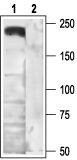
- Western blot analysis of rat brain membranes: - 1. Anti-CACNA1B (CaV2.2) Antibody (#ACC-002), (1:200). 2. Anti-CACNA1B (CaV2.2) Antibody , preincubated with the control antigen.
Supportive validation
- Submitted by
- Invitrogen Antibodies (provider)
- Main image
- Experimental details
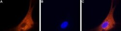
- Expression of CaV2.2 in rat DRG primary culture - Immunocytochemical staining of paraformaldehyde-fixed and permeabilized rat dorsal root ganglion (DRG) primary culture. A. Cells were stained using Anti-CACNA1B (CaV2.2) Antibody (#ACC-002), (1:200) followed by goat Anti-rabbit-AlexaFluor-555 secondary Antibody . B. Nuclear fluorescence staining of cells using the membrane-permeable DNA dye Hoechst 33342. C. Merged images of panels A and B.

Supportive validation
- Submitted by
- Invitrogen Antibodies (provider)
- Main image
- Experimental details
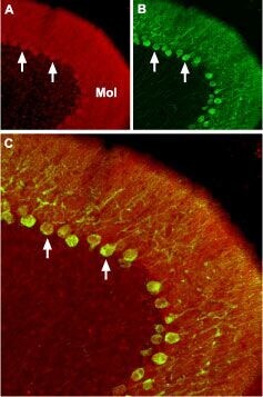
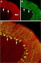
- Expression of CaV2.2 in mouse cerebellum - Immunohistochemical staining of mouse cerebellum with Anti-CACNA1B (CaV2.2) Antibody (#ACC-002), (1:100).A. CaV2.2 (red) appears in Purkinje cells (arrows) and is distributed diffusely in the molecular layer (Mol). B. Staining of Purkinje cells with mouse Anti-Calbindin 28K (green) demonstrates the restriction of CaV2.2 to cell bodies but not to dendrites in the molecular layer. C. Merged image of panels A and B.