Explore
 Validate
 Learn
Learn Western blot
 Immunocytochemistry
Antibody data
- Antibody Data
- Antigen structure
- References [2]
- Comments [0]
- Validations
- Western blot [4]
- Immunoprecipitation [1]
- Immunohistochemistry [1]
Submit
Validation data
Reference
Comment
Report error
- Product number
- NBP1-33021 - Provider product page

- Provider
- Novus Biologicals
- Proper citation
- Novus Cat#NBP1-33021, RRID:AB_2227785
- Product name
- Rabbit Polyclonal ATP6V1A Antibody
- Antibody type
- Polyclonal
- Description
- Immunogen affinity purified.
- Reactivity
- Human, Mouse
- Host
- Rabbit
- Isotype
- IgG
- Vial size
- 0.1 ml
- Storage
- Aliquot and store at -20C or -80C. Avoid freeze-thaw cycles.
Submitted references Clathrin coat controls synaptic vesicle acidification by blocking vacuolar ATPase activity.
Securin and separase modulate membrane traffic by affecting endosomal acidification.
Farsi Z, Gowrisankaran S, Krunic M, Rammner B, Woehler A, Lafer EM, Mim C, Jahn R, Milosevic I
eLife 2018 Apr 13;7
eLife 2018 Apr 13;7
Securin and separase modulate membrane traffic by affecting endosomal acidification.
Bacac M, Fusco C, Planche A, Santodomingo J, Demaurex N, Leemann-Zakaryan R, Provero P, Stamenkovic I
Traffic (Copenhagen, Denmark) 2011 May;12(5):615-26
Traffic (Copenhagen, Denmark) 2011 May;12(5):615-26
No comments: Submit comment
Supportive validation
- Submitted by
- Novus Biologicals (provider)
- Main image

- Experimental details
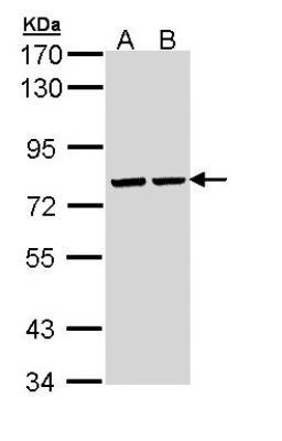
- Western Blot: ATP6V1A Antibody [NBP1-33021] - Sample (30 ug of whole cell lysate) A: Hela B: Hep G2 7. 5% SDS PAGE; antibody diluted at 1:10000.
- Submitted by
- Novus Biologicals (provider)
- Main image
- Experimental details
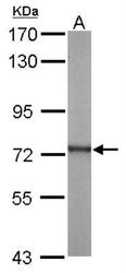
- Western Blot: ATP6V1A Antibody [NBP1-33021] - Sample (20 ug of whole cell lysate) A: mouse brain 7.5% SDS PAGE; antibody diluted at 1:5000.

- Submitted by
- Novus Biologicals (provider)
- Main image

- Experimental details
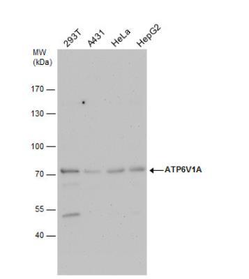
- Western Blot: ATP6V1A Antibody [NBP1-33021] - Various whole cell extracts (30ug) were separated by 7.5% SDS-PAGE, and the membrane was blotted with ATP6V1A antibody diluted by 1:10000.
- Submitted by
- Novus Biologicals (provider)
- Main image
- Experimental details
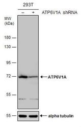
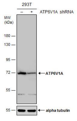
- Western Blot: ATP6V1A Antibody [NBP1-33021] - Non-transfected (-) and transfected (+) 293T whole cell extracts (30 ug) were separated by 7.5% SDS-PAGE, and the membrane was blotted with ATP6V1A antibody. HRP-conjugated anti-rabbit IgG antibody was used to detect the primary antibody.
Supportive validation
- Submitted by
- Novus Biologicals (provider)
- Main image
- Experimental details
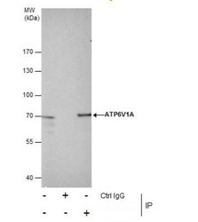
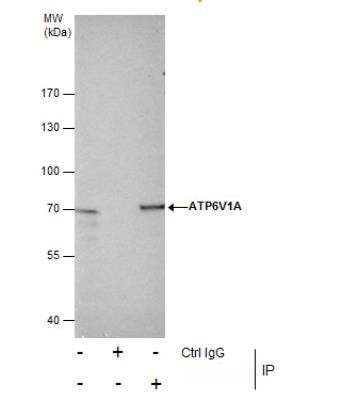
- Immunoprecipitation: ATP6V1A Antibody [NBP1-33021] - ATP6V1A Antibody [NBP1-33021] - Immunoprecipitation of ATP6V1A protein from HeLa whole cell extracts using 5 ug of ATP6V1A antibody. Western blot analysis was performed using ATP6V1A antibody EasyBlot anti-Rabbit IgG was used as a secondary reagent.
Supportive validation
- Submitted by
- Novus Biologicals (provider)
- Main image
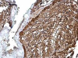
- Experimental details
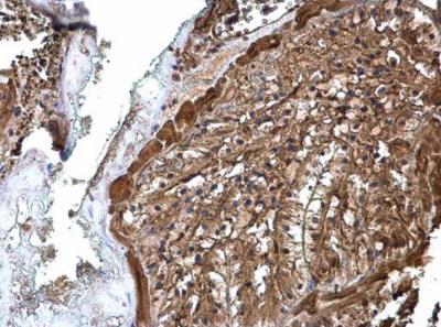
- Immunohistochemistry: ATP6V1A Antibody [NBP1-33021] - Mouse prostate ATP6V1A antibody diluted at 1:500.