Explore
 Validate
 Learn
Learn Western blot
Antibody data
- Antibody Data
- Antigen structure
- References [0]
- Comments [0]
- Validations
- Western blot [3]
- Immunocytochemistry [3]
- Immunohistochemistry [1]
- Flow cytometry [1]
Submit
Validation data
Reference
Comment
Report error
- Product number
- PA5-109274 - Provider product page

- Provider
- Invitrogen Antibodies
- Product name
- GPX4 Polyclonal Antibody
- Antibody type
- Polyclonal
- Antigen
- Recombinant full-length protein
- Reactivity
- Human
- Host
- Rabbit
- Isotype
- IgG
- Vial size
- 100 µL
- Concentration
- 1 mg/mL
- Storage
- -20° C, Avoid Freeze/Thaw Cycles, store in dark
No comments: Submit comment
Supportive validation
- Submitted by
- Invitrogen Antibodies (provider)
- Main image

- Experimental details
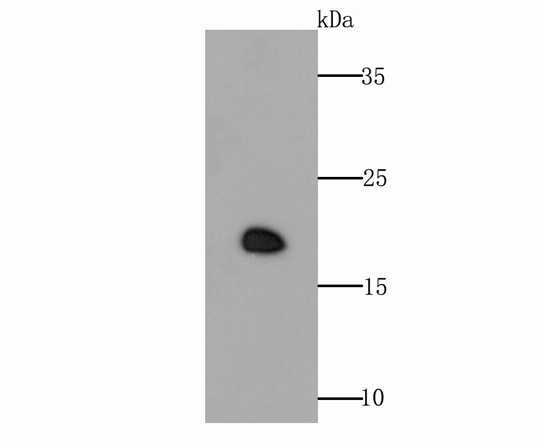
- Western blot analysis of GPX4 in rat testis tissue lysate. Samples were incubated with GPX4 polyclonal antibody (Product # PA5-109274), at a dilution of 1:500.

- Submitted by
- Invitrogen Antibodies (provider)
- Main image

- Experimental details
- Western blot analysis of GPX4 in rat testis tissue lysate. Samples were incubated with GPX4 polyclonal antibody (Product # PA5-109274), at a dilution of 1:500.
- Submitted by
- Invitrogen Antibodies (provider)
- Main image
- Experimental details
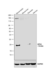
- Western blot was performed using Anti-GPX4 Polyclonal Antibody (Product # PA5-109274) and a 19 kDa band corresponding to GPX4 was observed in mouse and rat testes as opposed to mouse and rat liver tissues, as reported (doi: 10.1074/jbc.M109.016139). Tissue extracts (30 µg lysate) of Mouse Testis (Lane 1), Mouse Liver (Lane 2), Rat Testis (Lane 3), Rat Liver (Lane 4) were electrophoresed using NuPAGE™ 4-12% Bis-Tris Protein Gel (Product # NP0322BOX). Resolved proteins were then transferred onto a nitrocellulose membrane (Product # IB23001) by iBlot® 2 Dry Blotting System (Product # IB21001). The blot was probed with the primary antibody (1:1000 dilution) and detected by chemiluminescence with Goat anti-Rabbit IgG (H+L) Superclonal™ Recombinant Secondary Antibody, HRP (Product # A27036,1:20,000 dilution) using the iBright™ FL1500 Imaging System (Product # A44115). Chemiluminescent detection was performed using SuperSignal™ West Pico PLUS Chemiluminescent Substrate (Product # 34580).

Supportive validation
- Submitted by
- Invitrogen Antibodies (provider)
- Main image

- Experimental details
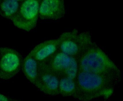
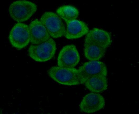
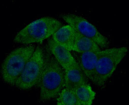
- Immunofluorescent analysis of GPX4 in MCF-7 cells (green). Samples were fixed in paraformaldehyde and permeabilised with 0.25% Triton X100/PBS, incubated with GPX4 polyclonal antibody (Product # PA5-109274), followed by DAPI (blue).
- Submitted by
- Invitrogen Antibodies (provider)
- Main image

- Experimental details
- Immunofluorescent analysis of GPX4 in LOVO cells (green). Samples were fixed in paraformaldehyde and permeabilised with 0.25% Triton X100/PBS, incubated with GPX4 polyclonal antibody (Product # PA5-109274), followed by DAPI (blue).
- Submitted by
- Invitrogen Antibodies (provider)
- Main image

- Experimental details
- Immunofluorescent analysis of GPX4 in HepG2 cells (green). Samples were fixed in paraformaldehyde and permeabilised with 0.25% Triton X100/PBS, incubated with GPX4 polyclonal antibody (Product # PA5-109274), followed by DAPI (blue).
Supportive validation
- Submitted by
- Invitrogen Antibodies (provider)
- Main image
- Experimental details
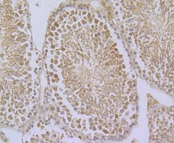
- Immunohistochemistry analysis of GPX4 in paraffin-embedded mouse testis tissue. Samples were incubated with GPX4 polyclonal antibody (Product # PA5-109274), and followed by hematoxylin.

Supportive validation
- Submitted by
- Invitrogen Antibodies (provider)
- Main image
- Experimental details
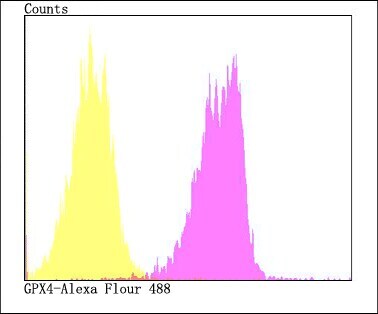
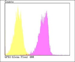
- Flow cytometry of GPX4 in HepG2 cells (fuchsia) compared with an unlabelled control (cells without incubation with primary antibody; yellow). Samples were incubated with GPX4 polyclonal antibody (Product # PA5-109274) at a dilution of 1:100, followed by Alexa Fluor 488-conjugated goat anti-rabbit IgG.