Explore
 Validate
 Learn
Learn Western blot
 ELISA
ELISAAntibody data
- Antibody Data
- Antigen structure
- References [0]
- Comments [0]
- Validations
- Western blot [1]
- Immunocytochemistry [1]
- Immunohistochemistry [1]
Submit
Validation data
Reference
Comment
Report error
- Product number
- MA5-27661 - Provider product page

- Provider
- Invitrogen Antibodies
- Product name
- SOD3 Monoclonal Antibody (4GG11G6)
- Antibody type
- Monoclonal
- Antigen
- Purifed from natural sources
- Description
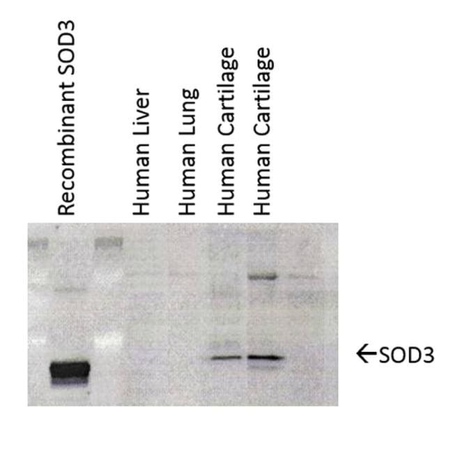
- 1 µg/mL of MA5-27661 was sufficient for detection of EC-SOD in 20 µg of human cartilage lysate by colorimetric immunoblot analysis using Goat anti-mouse IgG:HRP as the secondary antibody.|Detects extracellular SOD approximately 35kDa.
- Reactivity
- Human, Mouse, Rat, Guinea Pig
- Host
- Mouse
- Isotype
- IgG
- Antibody clone number
- 4GG11G6
- Vial size
- 100 µg
- Concentration
- 1 mg/mL
- Storage
- -20°C
No comments: Submit comment
Supportive validation
- Submitted by
- Invitrogen Antibodies (provider)
- Main image

- Experimental details
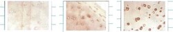
- Western blot analysis of SOD3 in human cartilage lysates. The sample was incubated with SOD3 monoclonal antibody (Product # MA5-27661) using a dilution of 1:1000. Samples were arranged as follows: Left) Control, Middle) Young cartilage, Right) Cartilage sample with osteoarthritis.
Supportive validation
- Submitted by
- Invitrogen Antibodies (provider)
- Main image

- Experimental details
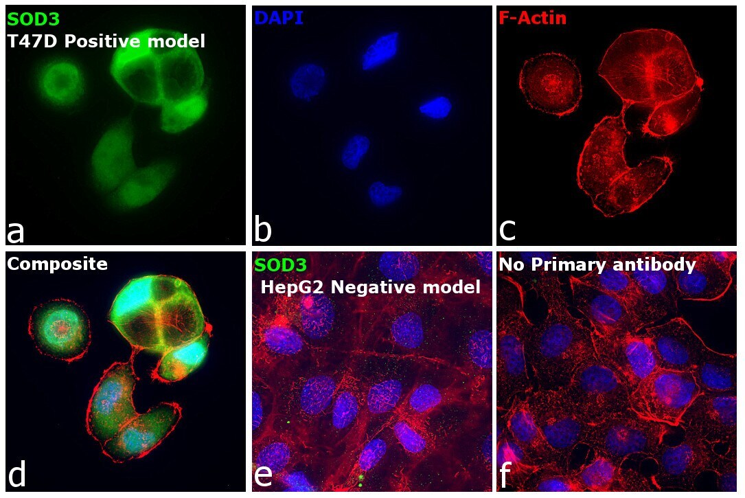
- Immunofluorescence analysis of Extracellular superoxide dismutase [Cu-Zn] was performed using 70% confluent log phase T-47D and Hep G2 cells. The cells were fixed with 4% paraformaldehyde for 10 minutes, permeabilized with 0.1% Triton™ X-100 for 15 minutes, and blocked with 2% BSA for 45 minutes at room temperature. The cells were labeled with SOD3 Monoclonal Antibody (4GG11G6) (Product # MA5-27661) at 1:100 in 0.1% BSA, incubated at 4 degree celsius overnight and then labeled with Donkey anti-Mouse IgG (H+L) Highly Cross-Adsorbed Secondary Antibody, Alexa Fluor Plus 488 (Product # A32766), (1:2000 dilution), for 45 minutes at room temperature (Panel a: Green). Nuclei (Panel b:Blue) were stained with ProLong™ Diamond Antifade Mountant with DAPI (Product # P36962). F-actin (Panel c: Red) was stained with Rhodamine Phalloidin (Product # R415, 1:300). Panel d represents the merged image showing Cytoplasmic localization in T-47D but not in Hep G2 (Panel e) which is reported to be low to negative for SOD3 expression. Panel f represents control cells with no primary antibody to assess background. The images were captured at 60X magnification.
Supportive validation
- Submitted by
- Invitrogen Antibodies (provider)
- Main image
- Experimental details
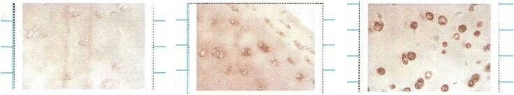
- Immunofluorescent analysis of SOD3 in mouse human cartilage. Sample was incubated with SOD3 monoclonal antibody (Product # MA5-27661) using a dilution of 1:1000. Images are shown as follows: Left: control, middle: young cartilage, right: cartilage with osteoarthritis.