 Explore
 Validate
 Learn
Learn Western blot
Antibody data
- Antibody Data
- Antigen structure
- References [1]
- Comments [0]
- Validations
- Western blot [1]
- Immunohistochemistry [1]
Submit
Validation data
Reference
Comment
Report error
- Product number
- AF5699 - Provider product page

- Provider
- R&D Systems
- Product name
- Human/Mouse SorLA Antibody
- Antibody type
- Polyclonal
- Description
- Antigen Affinity-purified. Detects human and mouse SorLA in direct ELISAs and Western blots.
- Reactivity
- Human, Mouse
- Host
- Sheep
- Conjugate
- Unconjugated
- Antigen sequence
Q92673- Isotype
- IgG
- Vial size
- 100 ug
- Concentration
- LYOPH
- Storage
- Use a manual defrost freezer and avoid repeated freeze-thaw cycles. 12 months from date of receipt, -20 to -70 °C as supplied. 1 month, 2 to 8 °C under sterile conditions after reconstitution. 6 months, -20 to -70 °C under sterile conditions after reconstitution.
Submitted references Prions amplify through degradation of the VPS10P sorting receptor sortilin.
Uchiyama K, Tomita M, Yano M, Chida J, Hara H, Das NR, Nykjaer A, Sakaguchi S
PLoS pathogens 2017 Jun;13(6):e1006470
PLoS pathogens 2017 Jun;13(6):e1006470
No comments: Submit comment
Supportive validation
- Submitted by
- R&D Systems (provider)
- Main image

- Experimental details
- Detection of Mouse SorLA by Western Blot. Western blot shows lysates of Neuro-2A mouse neuroblastoma cell line. PVDF Membrane was probed with 1 µg/mL of Sheep Anti-Human SorLA Antigen Affinity-purified Polyclonal Antibody (Catalog # AF5699) followed by HRP-conjugated Anti-Sheep IgG Secondary Antibody (Catalog # HAF016). A specific band was detected for SorLA at approximately 330 kDa (as indicated). This experiment was conducted under reducing conditions and using Immunoblot Buffer Group 1.

Supportive validation
- Submitted by
- R&D Systems (provider)
- Main image
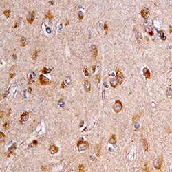
- Experimental details
- SorLA in Human Brain. SorLA was detected in immersion fixed paraffin-embedded sections of human brain using Sheep Anti-Human/Mouse SorLA Antigen Affinity-purified Polyclonal Antibody (Catalog # AF5699) at 15 µg/mL overnight at 4 °C. Tissue was stained using the Anti-Sheep HRP-DAB Cell & Tissue Staining Kit (brown; Catalog # CTS019) and counterstained with hematoxylin (blue). Specific staining was localized to neurons. View our protocol for Chromogenic IHC Staining of Paraffin-embedded Tissue Sections.
