 Explore
 Validate
 Learn
Learn Western blot
Antibody data
- Antibody Data
- Antigen structure
- References [1]
- Comments [0]
- Validations
- Western blot [2]
- Immunocytochemistry [1]
- Immunohistochemistry [2]
- Other assay [2]
Submit
Validation data
Reference
Comment
Report error
- Product number
- PA5-31113 - Provider product page

- Provider
- Invitrogen Antibodies
- Product name
- FSTL1 Polyclonal Antibody
- Antibody type
- Polyclonal
- Antigen
- Recombinant protein fragment
- Description
- Recommended positive controls: 293T, Jurkat.
- Concentration
- 1 mg/mL
Submitted references MicroRNA-27a Inhibits Cell Migration and Invasion of Fibroblast-Like Synoviocytes by Targeting Follistatin-Like Protein 1 in Rheumatoid Arthritis.
Shi DL, Shi GR, Xie J, Du XZ, Yang H
Molecules and cells 2016 Aug 31;39(8):611-8
Molecules and cells 2016 Aug 31;39(8):611-8
No comments: Submit comment
Supportive validation
- Submitted by
- Invitrogen Antibodies (provider)
- Main image

- Experimental details
- Western Blot using FSTL1 Polyclonal Antibody (Product # PA5-31113). Sample (30 µg of whole cell lysate). Lane A: Jurkat. 10% SDS PAGE. FSTL1 Polyclonal Antibody (Product # PA5-31113) diluted at 1:1,000.

- Submitted by
- Invitrogen Antibodies (provider)
- Main image

- Experimental details
- Western blot was performed using Anti-FSTL1 Polyclonal Antibody(Product # PA5-31113) and a 50kDa band corresponding to FSTL1 was observed across cell lines tested and tissue extracts tested except Mouse Liver which is reported to be negative. The expression of protein is also reported to be low in Caco-2 compared to other cell model. A faint non specific band around 18kDa was also observed. Whole cell extracts (30 µg lysate) of A549 (Lane 1), Caco-2 (Lane 2), HeLa (Lane 3), U-2 OS (Lane 4), HCT 116 (Lane 5), PC-12 (Lane 6) and tissue extracts of Mouse Liver (Lane 7), Mouse Brown Adipose (Lane 8) and Mouse Lung (Lane 9) were electrophoresed using NuPAGE™ 4-12% Bis-Tris Protein Gel (Product # NP0322BOX). Resolved proteins were then transferred onto a Nitrocellulose membrane (Product # IB23001) by iBlot® 2 Dry Blotting System (Product # IB21001). The blot was probed with the primary antibody (1:3000 dilution) and detected by chemiluminescence with Goat anti-Rabbit IgG (H+L) Superclonal™ Recombinant Secondary Antibody, HRP (Product # A27036, 1:4000 dilution) using the iBright FL 1000 (Product # A32752). Chemiluminescent detection was performed using Novex® ECL Chemiluminescent Substrate Reagent Kit (Product # WP20005).

Supportive validation
- Submitted by
- Invitrogen Antibodies (provider)
- Main image

- Experimental details
- Immunofluorescence analysis of FSTL1 was performed using 70% confluent log phase A549 cells. The cells were fixed with 4% paraformaldehyde for 10 minutes, permeabilized with 0.1% Triton™ X-100 for 15 minutes, and blocked with 2% BSA for 45 minutes at room temperature. The cells were labeled with FSTL1 Polyclonal Antibody (Product # PA5-31113) at 5 µg/mL in 0.1% BSA, incubated at 4 degree celsius overnight and then labeled with Goat anti-Rabbit IgG (H+L) Highly Cross-Adsorbed Secondary Antibody, Alexa Fluor Plus 488 (Product # A32731), (1:2000), for 45 minutes at room temperature (Panel a: Green). Nuclei (Panel b:Blue) were stained with ProLong™ Diamond Antifade Mountant with DAPI (Product # P36962). F-actin (Panel c: Red) was stained with Rhodamine Phalloidin (Product # R415, 1:300). Panel d represents the merged image showing cytoplasmic localization. Panel e represents control cells with no primary antibody to assess background. The images were captured at 60X magnification.

Supportive validation
- Submitted by
- Invitrogen Antibodies (provider)
- Main image

- Experimental details
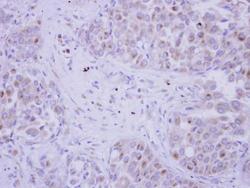
- FSTL1 Polyclonal Antibody detects FSTL1 protein at cytosol on human breast carcinoma by immunohistochemical analysis. Sample: Paraffin-embedded human breast carcinoma. FSTL1 Polyclonal Antibody (Product # PA5-31113) dilution: 1:250. Scale bar = 100 µm. Antigen Retrieval: EDTA based buffer, pH 8.0, 15 min.

- Submitted by
- Invitrogen Antibodies (provider)
- Main image
- Experimental details
- Immunohistochemical analysis of paraffin-embedded human breast cancer, using FSTL1 (Product # PA5-31113) antibody at 1:250 dilution. Antigen Retrieval: EDTA based buffer, pH 8.0, 15 min.

Supportive validation
- Submitted by
- Invitrogen Antibodies (provider)
- Main image

- Experimental details
- Fig. 1. Expression of miR-27a and FSTL1 in RA serum, synovial tissue, and FLS. (A) The serum expression of FSTL1 of RA patients was validated by ELISA, and its level in synovial tissue and FLS was validated by western blot and qRT-PCR. (B) The expression of miR-27a was validated in serum, synovial tissue, and FLS of RA patients by qRT-PCR. ** p < 0.01, versus control group.

- Submitted by
- Invitrogen Antibodies (provider)
- Main image

- Experimental details
- Fig. 4. Identification of FSTL1 as a target of miR-27a. (A) A luciferase reporter with either the wild-type or mutant sequences of FSTL1 3'UTR was constructed according to the sequence of the miR-27a binding site within the 3'UTR of human FSTL1, using the pGL3 vector. (B) For the luciferase reporter assay, FLS were co-transfected with the luciferase reporter vectors and control mimic, miR-27a mimic, control inhibitor, or miR-27a inhibitor. Relative luciferase activity in RA-FLS was measured after a 48-h transfection. (C) Western blot analysis was used to evaluate the expression of FSTL1 in RA-FLS after transfection with miR-27a mimic or miR-27a inhibitor. * p < 0.01 versus control group.
