 Explore
 Validate
 Learn
Learn Western blot
Antibody data
- Antibody Data
- Antigen structure
- References [3]
- Comments [0]
- Validations
- Western blot [2]
- Immunohistochemistry [2]
Submit
Validation data
Reference
Comment
Report error
- Product number
- AF1970 - Provider product page

- Provider
- R&D Systems
- Product name
- Human Thioredoxin-1 Antibody
- Antibody type
- Polyclonal
- Description
- Antigen Affinity-purified. Detects human Thioredoxin-1 in direct ELISAs and Western blots. In direct ELISAs, less than 25% cross-reactivity with recombinant mouse Thioredoxin-1 is observed.
- Reactivity
- Human
- Host
- Goat
- Conjugate
- Unconjugated
- Antigen sequence
P10599- Isotype
- IgG
- Vial size
- 100 ug
- Concentration
- LYOPH
- Storage
- Use a manual defrost freezer and avoid repeated freeze-thaw cycles. 12 months from date of receipt, -20 to -70 °C as supplied. 1 month, 2 to 8 °C under sterile conditions after reconstitution. 6 months, -20 to -70 °C under sterile conditions after reconstitution.
Submitted references Thioredoxin increases exocytosis by denitrosylating N-ethylmaleimide-sensitive factor.
Thioredoxin-related mechanisms in hyperoxic lung injury in mice.
Thioredoxin is required for S-nitrosation of procaspase-3 and the inhibition of apoptosis in Jurkat cells.
Ito T, Yamakuchi M, Lowenstein CJ
The Journal of biological chemistry 2011 Apr 1;286(13):11179-84
The Journal of biological chemistry 2011 Apr 1;286(13):11179-84
Thioredoxin-related mechanisms in hyperoxic lung injury in mice.
Tipple TE, Welty SE, Rogers LK, Hansen TN, Choi YE, Kehrer JP, Smith CV
American journal of respiratory cell and molecular biology 2007 Oct;37(4):405-13
American journal of respiratory cell and molecular biology 2007 Oct;37(4):405-13
Thioredoxin is required for S-nitrosation of procaspase-3 and the inhibition of apoptosis in Jurkat cells.
Mitchell DA, Morton SU, Fernhoff NB, Marletta MA
Proceedings of the National Academy of Sciences of the United States of America 2007 Jul 10;104(28):11609-14
Proceedings of the National Academy of Sciences of the United States of America 2007 Jul 10;104(28):11609-14
No comments: Submit comment
Supportive validation
- Submitted by
- R&D Systems (provider)
- Main image

- Experimental details
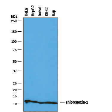
- Detection of Human Thioredoxin-1 by Western Blot. Western blot shows lysates of HeLa human cervical epithelial carcinoma cell line, HepG2 human hepatocellular carcinoma cell line, Jurkat human acute T cell leukemia cell line, K562 human chronic myelogenous leukemia cell line, and Raji human Burkitt's lymphoma cell line. PVDF membrane was probed with 0.1 µg/mL of Goat Anti-Human Thioredoxin-1 Antigen Affinity-purified Polyclonal Antibody (Catalog # AF1970) followed by HRP-conjugated Anti-Goat IgG Secondary Antibody (Catalog # HAF019). A specific band was detected for Thioredoxin-1 at approximately 12 kDa (as indicated). This experiment was conducted under reducing conditions and using Immunoblot Buffer Group 1.
- Submitted by
- R&D Systems (provider)
- Main image
- Experimental details
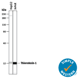
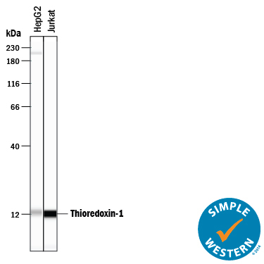
- Detection of Human Thioredoxin-1 by Simple WesternTM. Simple Western lane view shows lysates of HepG2 human hepatocellular carcinoma cell line and Jurkat human acute T cell leukemia cell line, loaded at 0.2 mg/mL. A specific band was detected for Thioredoxin-1 at approximately 12 kDa (as indicated) using 1 µg/mL of Goat Anti-Human Thioredoxin-1 Antigen Affinity-purified Polyclonal Antibody (Catalog # AF1970) followed by 1:50 dilution of HRP-conjugated Anti-Goat IgG Secondary Antibody (Catalog # HAF109). This experiment was conducted under reducing conditions and using the 12-230 kDa separation system.
Supportive validation
- Submitted by
- R&D Systems (provider)
- Main image
- Experimental details
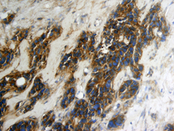
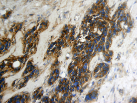
- Thioredoxin-1 in Human Breast Cancer Tissue. Thioredoxin-1 was detected in immersion fixed paraffin-embedded sections of human breast cancer tissue using Goat Anti-Human Thioredoxin-1 Antigen Affinity-purified Polyclonal Antibody (Catalog # AF1970) at 15 µg/mL overnight at 4 °C. Tissue was stained using the Anti-Goat HRP-DAB Cell & Tissue Staining Kit (brown; Catalog # CTS008) and counterstained with hematoxylin (blue). View our protocol for Chromogenic IHC Staining of Paraffin-embedded Tissue Sections.
- Submitted by
- R&D Systems (provider)
- Main image
- Experimental details
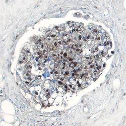
- Thioredoxin-1 in Human Breast Cancer Tissue. Thioredoxin-1 was detected in immersion fixed paraffin-embedded sections of human breast cancer tissue using Goat Anti-Human Thioredoxin-1 Antigen Affinity-purified Polyclonal Antibody (Catalog # AF1970) at 5 µg/mL overnight at 4 °C. Tissue was stained using the Anti-Goat HRP-DAB Cell & Tissue Staining Kit (brown; Catalog # CTS008) and counterstained with hematoxylin (blue). View our protocol for Chromogenic IHC Staining of Paraffin-embedded Tissue Sections.
