Explore
 Validate
 Learn
Learn Western blot
Antibody data
- Antibody Data
- Antigen structure
- References [0]
- Comments [0]
- Validations
- Western blot [1]
- Immunocytochemistry [2]
- Immunohistochemistry [1]
- Flow cytometry [1]
Submit
Validation data
Reference
Comment
Report error
- Product number
- TA325114 - Provider product page

- Provider
- OriGene
- Product name
- Rabbit polyclonal ZNF202 Antibody (Center)
- Antibody type
- Polyclonal
- Description
- Rabbit polyclonal ZNF202 Antibody (Center)
- Host
- Rabbit
- Conjugate
- Unconjugated
- Epitope
- ZNF202
- Isotype
- IgG
- Antibody clone number
- NULL
- Vial size
- 400 µl
- Concentration
- 0.48 mg/ml
No comments: Submit comment
Supportive validation
- Submitted by
- OriGene (provider)
- Main image
- Experimental details
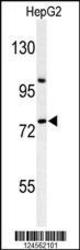
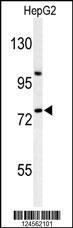
- Western blot analysis of ZNF202 Antibody (Center) (Cat. #TA325114) in HepG2 cell line lysates (35ug/lane). ZNF202 (arrow) was detected using the purified Pab.
- Validation comment
- WB
Supportive validation
- Submitted by
- OriGene (provider)
- Main image

- Experimental details
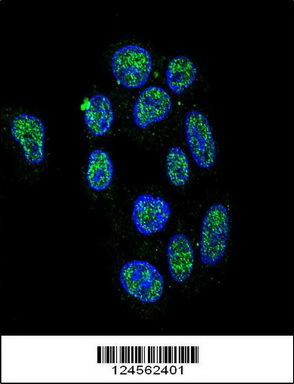
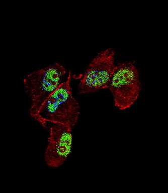
- Confocal immunofluorescent analysis of ZNF202 Antibody (Center) (Cat#TA325114) with HepG2 cell followed by Alexa Fluor 488-conjugated goat anti-rabbit lgG (green). DAPI was used to stain the cell nuclear (blue).
- Validation comment
- IF
- Submitted by
- OriGene (provider)
- Main image

- Experimental details
- IF image of MDA-MB231 cell stained with ZNF202 Antibody (Center)(Cat#TA325114). MDA-MB231 cells were incubated with ZNF202 primary antibody (1:25, 1 h at 37?). For secondary antibody, Alexa Fluor? 488 conjugated donkey anti-rabbit antibody (green) was used (1:400).Cytoplasmic actin was counterstained with Alexa Fluor? 555 (red) conjugated Phalloidin (7 units/ml). Nuclei were counterstained with DAPI (blue) .ZNF202 immunoreactivity is localized to nucleus significantly.
- Validation comment
- IF
Supportive validation
- Submitted by
- OriGene (provider)
- Main image
- Experimental details
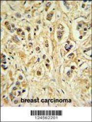
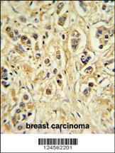
- ZNF202 Antibody (Center) (Cat. #TA325114) IHC analysis in formalin fixed and paraffin embedded breast carcinoma followed by peroxidase conjugation of the secondary antibody and DAB staining. This data demonstrates the use of the ZNF202 Antibody (Center) for immunohistochemistry. Clinical relevance has not been evaluated.
- Validation comment
- IHC
Supportive validation
- Submitted by
- OriGene (provider)
- Main image
- Experimental details
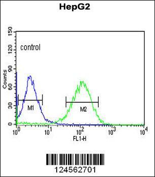
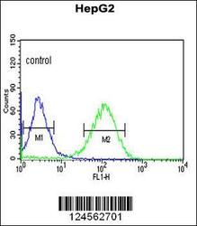
- ZNF202 Antibody (Center) (Cat. #TA325114) flow cytometric analysis of HepG2 cells (right histogram) compared to a negative control cell (left histogram).FITC-conjugated goat-anti-rabbit secondary antibodies were used for the analysis.
- Validation comment
- FC