Antibody data
- Antibody Data
- Antigen structure
- References [2]
- Comments [0]
- Validations
- Western blot [2]
Submit
Validation data
Reference
Comment
Report error
- Product number
- R1598PS - Provider product page

- Provider
- Acris Antibodies GmbH
- Product name
- anti Plasminogen / PLG
- Antibody type
- Polyclonal
- Antigen
- Plasminogen from human plasma
- Reactivity
- Human
- Host
- Goat
- Vial size
- 0.1 mg
- Concentration
- 1.0 mg/ml (by UV absorbance at 280 nm)
Submitted references Molecular characterization of the interaction of Borrelia parkeri and Borrelia turicatae with human complement regulators.
Gpm1p is a factor H-, FHL-1-, and plasminogen-binding surface protein of Candida albicans.
Schott M, Grosskinsky S, Brenner C, Kraiczy P, Wallich R
Infection and immunity 2010 May;78(5):2199-208
Infection and immunity 2010 May;78(5):2199-208
Gpm1p is a factor H-, FHL-1-, and plasminogen-binding surface protein of Candida albicans.
Poltermann S, Kunert A, von der Heide M, Eck R, Hartmann A, Zipfel PF
The Journal of biological chemistry 2007 Dec 28;282(52):37537-44
The Journal of biological chemistry 2007 Dec 28;282(52):37537-44
No comments: Submit comment
Supportive validation
- Submitted by
- Acris Antibodies GmbH (provider)
- Main image

- Experimental details
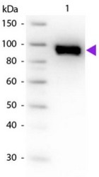
- Western blot using Plasminogen primary antibody Cat.-No. R1598P. Lane 1: Plasminogen. Load: 50 ng per lane. Primary antibody: Plasminogen primary antibody at 1/1,000 overnight at 4°C. Secondary antibody: Peroxidase goat secondary antibody at 1/40,000 for 60 min at RT. Blocking for 30 min at RT. Predicted/observed size: 91 kDa for Plasminogen. Other band(s): None.
- Submitted by
- Acris Antibodies GmbH (provider)
- Main image

- Experimental details
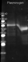
- Plasminogen antibody Cat.-No. R1598P, lot 6571) was used to detect plasminogen under reducing (R) and non-reducing (NR) conditions. Reduced samples of purified target proteins contained 4% BME and were boiled for 5 minutes. Samples of ~1µg of protein per lane were run by SDS-PAGE. Protein was transferred to nitrocellulose and probed with 1/3000 dilution of primary antibody. Detection shown was using Dylight 649 conjugated donkey anti goat for 1 hr RT. Images were collected using the BioRad VersaDoc System.
 Explore
 Validate
 Learn
Learn Western blot
 ELISA
ELISA