Explore
 Validate
 Learn
Learn Western blot
Antibody data
- Antibody Data
- Antigen structure
- References [4]
- Comments [0]
- Validations
- Western blot [5]
- Immunocytochemistry [1]
- Immunohistochemistry [3]
- Other assay [2]
Submit
Validation data
Reference
Comment
Report error
- Product number
- PA5-34677 - Provider product page

- Provider
- Invitrogen Antibodies
- Product name
- Plasminogen Polyclonal Antibody
- Antibody type
- Polyclonal
- Antigen
- Recombinant protein fragment
- Description
- Recommended positive controls: Jurkat, Raji, K562, BCL-1, PC-12, Rat2, human plasma, mouse plasma, rat plasma.
- Concentration
- 0.18 mg/mL
Submitted references Schistosoma mansoni phosphoglycerate mutase: a glycolytic ectoenzyme with thrombolytic potential.
Longitudinal proteomics study of serum changes after allogeneic HSCT reveals potential markers of metabolic complications related to aGvHD.
PLG inhibits Hippo signaling pathway through SRC in the hepatitis B virus-induced hepatocellular-carcinoma progression.
Schistosoma mansoni glyceraldehyde-3-phosphate dehydrogenase enhances formation of the blood-clot lysis protein plasmin.
Pirovich DB, Da'dara AA, Skelly PJ
Parasite (Paris, France) 2022;29:41
Parasite (Paris, France) 2022;29:41
Longitudinal proteomics study of serum changes after allogeneic HSCT reveals potential markers of metabolic complications related to aGvHD.
Wong SY, Kato S, Rodenburg F, Tojo A, Hayashi N
Scientific reports 2022 Aug 17;12(1):14002
Scientific reports 2022 Aug 17;12(1):14002
PLG inhibits Hippo signaling pathway through SRC in the hepatitis B virus-induced hepatocellular-carcinoma progression.
Hu ZG, Chen YB, Huang M, Tu JB, Tu SJ, Pan YJ, Chen XL, He SQ
American journal of translational research 2021;13(2):515-531
American journal of translational research 2021;13(2):515-531
Schistosoma mansoni glyceraldehyde-3-phosphate dehydrogenase enhances formation of the blood-clot lysis protein plasmin.
Pirovich DB, Da'dara AA, Skelly PJ
Biology open 2020 Mar 24;9(3)
Biology open 2020 Mar 24;9(3)
No comments: Submit comment
Supportive validation
- Submitted by
- Invitrogen Antibodies (provider)
- Main image
- Experimental details
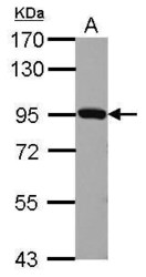
- Western Blot using Plasminogen Polyclonal Antibody (Product # PA5-34677). Sample (30 µg of whole cell lysate). Lane A: BCL-1. 7.5% SDS PAGE. Plasminogen Polyclonal Antibody (Product # PA5-34677) diluted at 1:1,000. The HRP-conjugated anti-rabbit IgG antibody was used to detect the primary antibody.

- Submitted by
- Invitrogen Antibodies (provider)
- Main image
- Experimental details
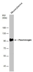
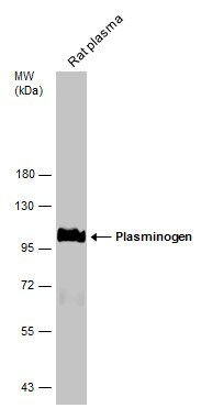
- Western Blot using Plasminogen Polyclonal Antibody (Product # PA5-34677). Mouse tissue extract (50 µg) was separated by 7.5% SDS-PAGE, and the membrane was blotted with Plasminogen Polyclonal Antibody (Product # PA5-34677) diluted at 1:500. The HRP-conjugated anti-rabbit IgG antibody was used to detect the primary antibody.
- Submitted by
- Invitrogen Antibodies (provider)
- Main image

- Experimental details
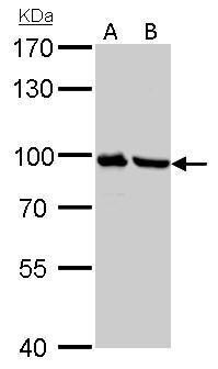
- Plasminogen Polyclonal Antibody detects PLG protein by western blot analysis. A. 30 µg PC-12 whole cell lysate/extract. B. 30 µg Rat2 whole cell lysate/extract.7.5% SDS-PAGE. Plasminogen Polyclonal Antibody (Product # PA5-34677) dilution: 1:1,000. The HRP-conjugated anti-rabbit IgG antibody was used to detect the primary antibody.
- Submitted by
- Invitrogen Antibodies (provider)
- Main image

- Experimental details
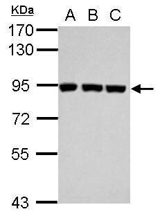
- Western Blot using Plasminogen Polyclonal Antibody (Product # PA5-34677). Sample (30 µg of whole cell lysate). Lane A: Jurkat. Lane B: Raji. Lane C: K562. 7.5% SDS PAGE. Plasminogen Polyclonal Antibody (Product # PA5-34677) diluted at 1:1,000. The HRP-conjugated anti-rabbit IgG antibody was used to detect the primary antibody.
- Submitted by
- Invitrogen Antibodies (provider)
- Main image
- Experimental details
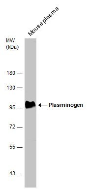
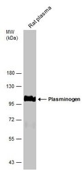
- Western Blot analysis of Plasminogen was performed by separating 50 µg of Rat tissue extracts by 7.5% SDS-PAGE. Proteins were transferred to a membrane and probed with a Plasminogen Polyclonal Antibody (Product # PA5-34677) at a dilution of 1:500. The HRP-conjugated anti-rabbit IgG antibody was used to detect the primary antibody.
Supportive validation
- Submitted by
- Invitrogen Antibodies (provider)
- Main image

- Experimental details
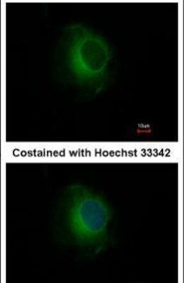
- Immunofluorescent analysis of Plasminogen in methanol-fixed HeLa cells using a Plasminogen polyclonal antibody (Product # PA5-34677) at a 1:500 dilution.
Supportive validation
- Submitted by
- Invitrogen Antibodies (provider)
- Main image

- Experimental details
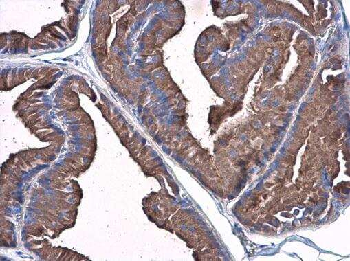
- Immunohistochemistry (Paraffin) analysis of Plasminogen was performed in paraffin-embedded rat prostate tissue using Plasminogen Polyclonal Antibody (Product # PA5-34677) at a dilution of 1:500.
- Submitted by
- Invitrogen Antibodies (provider)
- Main image
- Experimental details
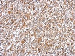
- Immunohistochemical analysis of paraffin-embedded C2C12 xenograft, using Plasminogen (Product # PA5-34677) antibody at 1:500 dilution. Antigen Retrieval: EDTA based buffer, pH 8.0, 15 min.
- Submitted by
- Invitrogen Antibodies (provider)
- Main image
- Experimental details
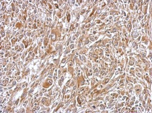
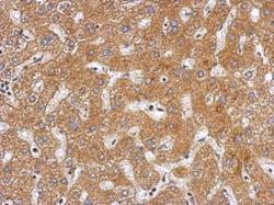
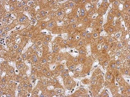
- Immunohistochemical analysis of paraffin-embedded human hepatoma, using Plasminogen (Product # PA5-34677) antibody at 1:500 dilution. Antigen Retrieval: EDTA based buffer, pH 8.0, 15 min.
Supportive validation
- Submitted by
- Invitrogen Antibodies (provider)
- Main image
- Experimental details
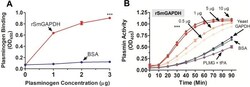
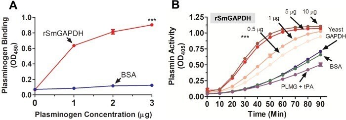
- Fig. 5. Recombinant SmGAPDH binds to plasminogen and enhances plasmin generation. (A) Plasminogen binding to rSmGAPDH (0.5 mug, red line) versus control protein BSA (0.5 mug, dark blue line) detected by ELISA (mean of triplicate OD 450 values+-s.e.m.). Significant difference between plasminogen binding to rSmGAPDH and BSA at all assay points are denoted by *** P
- Submitted by
- Invitrogen Antibodies (provider)
- Main image

- Experimental details
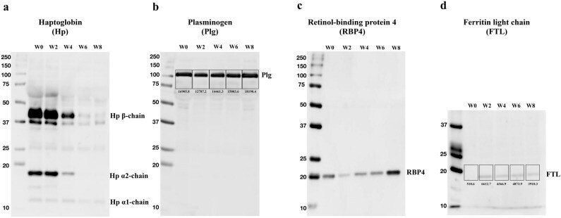
- Post-transplant serial changes in four serum proteins from Patient no.1. Serum samples collected over time from patient no.1 were subjected to western blotting analysis. Serum proteins of 10 ug was first separated by 8-12% SDS-PAGE and Precision Plus Protein WesternC Standard (Bio-Rad) was used as protein standard. Proteins were then transferred to a PVDF membrane and stained with antibodies. Primary antibodies were diluted at 2000-fold except for FTL (1000-fold). HRP Goat Anti-Rabbit IgG was used as secondary antibody in 2000-fold dilution except for RBP4 (10,000-fold). Signals of bands were quantified using ImageJ software.